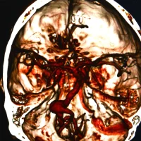
as shown in the lower image is associated with frailty and reduced lifespan in hip-fracture patients.")
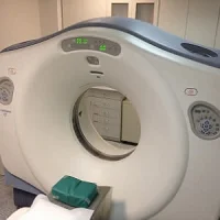
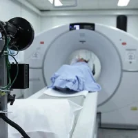
Findings from a new study conducted by radiologists at UC Davis and Wake Forest Bapist medical centres suggest that using computed tomography (CT) to evaluate muscle health may help identify optimal treatments for older patients who fall and break their hips. The study is published in the American Journal of Roentgenology.
This is the first study that links the use of imaging technology with survival after hip fractures. The study was condcuted with 300 participants at least 65 years of age and treated for fall-related injuries. All participants were suspected of breaking their hips. Patients received CTs to diagnose or rule out fracture. CTs were evaluated with additional measurements of size and density of the lumbar and thoracic muscle. Information was then compared with mortality data from the National Death Index.
Research findings showed that decreased core muscle that stabilises the spine was associated with decreased survival times after hip fractures. Patients with better core muscle had better survival rates.
Hip fractures are one of the most common causes of injury, hospitalisation and disability among older Americans. By using CT and by gathering information about muscle loss, doctors can determine the frailty level of a patient. This can in turn guide treatment decisions. If CTs show frailty in a patient, that patient would benefit more from a simpler surgery as compared to a patient with favourable life expectancy who could be treated with total hip arthroplasty.
As lead author Robert Boutin explains, the use of CT scans to evaluate muscles and hip bones can better help predict longevity and this in turn can help personalise treatment as per the patient's needs.
The researchers are hopeful that their findings will inspire additional studies in this area and would expand research that is focused on improving orthopaedic treatments for older patients.
Source: University of California - Davis Health System
Image Credit: Copyright UC Regents 2017
References:
Boutin, Robert D. et al. (2017) CT of Patients With Hip Fracture: Muscle Size and Attenuation Help Predict Mortality. American Journal of Roentgenology. doi/10.2214/AJR.16.17226
Latest Articles
CT scans, frailty, muscle health, hip fractures
Findings from a new study conducted by radiologists at UC Davis and Wake Forest Baptist medical centres suggest that using computed tomography (CT) to evaluate muscle health may help identify optimal treatments for older patients who fall and break their