Radiomics is a method that extracts detailed data, known as radiomic features, from medical images like CT and MRI. These features can reveal tissue characteristics at a microscopic level, potentially serving as biomarkers in oncology. They offer a non-invasive way to assess entire tumours and track changes over time. Radiomic features include shape, histogram-based, texture, and wavelet categories, each describing different aspects of image properties.
Lung cancer has been extensively studied using radiomics with CT and PET scans due to their widespread use in clinical settings. MRI, though offering superior soft tissue contrast and no radiation exposure, faces challenges in lung imaging due to low signal-to-noise ratios and movement artifacts. Despite these obstacles, some studies have explored MRI's potential in extracting radiomic features to predict treatment response and survival.
Comparing radiomic features between CT and MRI is complex because these imaging techniques capture different physical properties of tissues. However, understanding correlations between their features could reveal insights into lung cancer biology. To investigate this, researchers used LIFEx and PyRadiomics software to analyse features from CT and MRI scans, considering the impact of voxel resampling on their findings.
Overall, the study aimed to explore how well CT and MRI radiomic features correlate, potentially uncovering common biological aspects of lung cancer detectable by both imaging modalities.
Patient Characteristics and Imaging Protocol for Radiomic Analysis in Non-Small Cell Lung Cancer
From April 2021 to June 2023, thirty-five patients diagnosed with non-small cell lung cancer (NSCLC) were included. The cohort predominantly comprised males (74%) with a median age of 68 years. The histological distribution of NSCLC types included adenocarcinoma (37%), squamocellular carcinoma (34%), and poorly differentiated NSCLC (29%). Tumour sizes ranged from 2 cm to 15 m, corresponding to stage II to IV.
Exclusion criteria encompassed patients with MRI contraindications, prior treatment, or non-NSCLC lung tumours. All participants underwent thoracic CT using various scanners and parameters, followed by MRI using a 1.5-T system. MRI sequences included axial and coronal VIBE T1-weighted images post-contrast.
For image analysis, contrast-enhanced axial CT and T1-weighted MRI images were selected. Tumour segmentation was semi-automatically performed for CT and manually for MRI due to technological limitations. Segmentation accuracy was ensured by three radiologists, with complex cases reviewed collaboratively.
This methodology aimed to explore the correlation between radiomic features extracted from CT and MRI scans, focusing on their potential utility in characterizing NSCLC.
Radiomic Feature Reliability in CT and MRI: Implications for Cross-Modality Integration
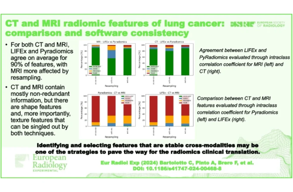
The study assessed agreement between radiomic features computed using LIFEx and PyRadiomics software across CT and MRI scans, employing three voxel resampling strategies. For MRI, reliability was generally high, with 95% of features showing excellent or good agreement without voxel resampling, 83% with internal resampling, and 91% with external resampling. Notably, poor or moderate reliability was observed for a small percentage of features, particularly Inverse Variance (GLCM) across all resampling methods.
In CT imaging, 92% of features exhibited excellent or good reliability without voxel resampling, slightly varying to 89% with internal resampling and 94% with external resampling. Poor or moderate reliability affected a small proportion of features, notably including GLCM Inverse Variance and several GLZLM features across all resampling strategies.
The comparison between CT and MRI features revealed limited agreement, particularly with PyRadiomics software where only 3% of features showed excellent or good reliability without voxel resampling, increasing to 11% with internal and 9% with external resampling. Conversely, LIFEx showed 5% reliability without resampling, with 9% internal and 11% external reliability. Notably, SHAPE features like Volume and Surface Area consistently demonstrated good/excellent reliability across all methods. Texture-based features such as NGTDM Busyness and Strength, and GLZLM ZP, also showed promising agreement under specific resampling conditions.
Overall, the study highlights the variability in reliability across different radiomic features extracted from CT and MRI scans using different software and voxel resampling methods, emphasizing the complexity and potential challenges in integrating radiomic analyses across imaging modalities.
Radiomic Feature Agreement Between CT and MRI: Insights into Methodological Variability and Cross-Modality Challenge
For MRI, the analysis revealed that at least 83% of features demonstrated good or excellent agreement (ICC ≥ 0.75) with each of the considered resampling methods. Specifically, the highest agreement was observed with original voxel dimensions (94.5% of features), while slightly lower agreement was found with internal resampling methods (83% of features). This discrepancy in agreement could potentially be attributed to differences in the resampling algorithms employed by LIFEx and PyRadiomics, which became more pronounced with internal resampling operations.
In contrast, CT images showed consistent reliability across different resampling methods, with at least 89% of features demonstrating good or excellent agreement. This stability in agreement was attributed to lower noise levels inherent in CT images compared to MRI, as well as the closer alignment of original voxel dimensions with isotropic sizes, minimizing the impact of resampling on feature extraction.
When comparing radiomic features extracted from CT and MRI scans, the study found that only around 10% of features exhibited good or excellent agreement between the two imaging modalities. Notably, features related to tumour shape (e.g., Volume, Surface Area) and certain texture features (e.g., NGTDM Busyness, Strength) showed the highest levels of agreement across both software packages and resampling methods. However, features such as GLCM Inverse Variance and several GLZLM features consistently demonstrated poor to moderate reliability across all resampling strategies, indicating challenges in their consistent extraction between CT and MRI.
Overall, the study underscored the importance of considering resampling methods in radiomic analyses, as these choices significantly influenced the agreement between LIFEx and PyRadiomics. The findings suggest that while there are shared radiomic features reflecting NSCLC biology across CT and MRI, achieving robust biomarkers requires careful methodological standardization and validation across larger cohorts and multi-centre studies.
Source & Image Credit: European Radiology Experimental

