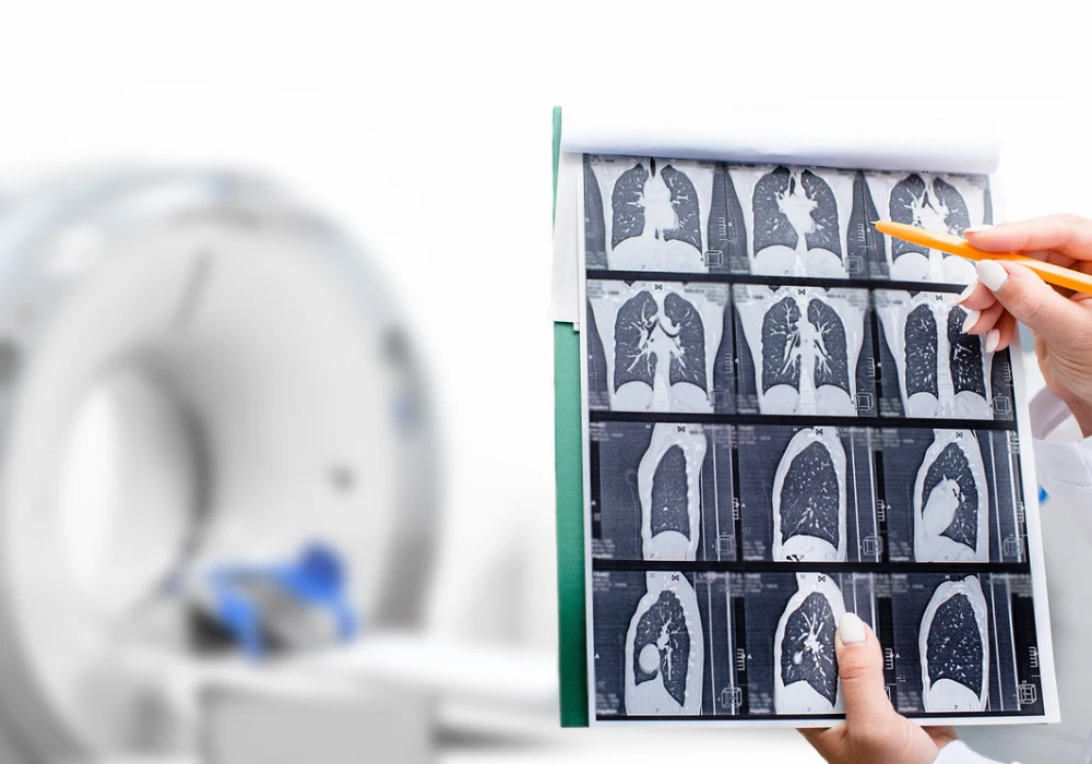
Coronary artery calcium (CAC) scoring is a valuable diagnostic tool for assessing cardiovascular risk, particularly in asymptomatic individuals. Traditionally, this assessment relies on electrocardiogram (ECG)-gated computed tomography (CT) scans to quantify calcified plaque burden in the coronary arteries. However, such scans are not always available in all clinical settings. Recent guidelines have encouraged the use of low-dose chest CT (LDCT), primarily conducted for lung cancer screening, to opportunistically assess CAC.
While manual scoring on LDCT can yield useful insights, it remains time-consuming and prone to variability. The advent of artificial intelligence, specifically deep learning (DL), has opened the door to automation in this field. A recent study published in European Radiology evaluated the performance of a fully automated DL-based coronary artery calcium scoring (DL-CACS) system across both ECG-gated calcium CT and non-gated LDCT modalities using multivendor datasets. It aimed to determine whether DL-CACS can provide accurate, efficient and clinically viable results that match or exceed manual methods.
Outstanding Accuracy in ECG-Gated CT Scans
The DL-CACS system performed with exceptional precision on ECG-gated calcium CT scans. In both the Seoul National University Hospital (SNUH) dataset and the public Stanford-COCA dataset, correlation metrics such as the intraclass correlation coefficient (ICC) and the coefficient of determination (R²) exceeded 0.99. These values indicate near-perfect alignment between the automated system and expert-generated reference scores. The categorical agreement, which assesses how well the model classifies patients into clinically relevant risk groups, also reached impressive levels, with weighted kappa values nearing or exceeding 0.97. The area under the receiver operating characteristic curve (AUROC), used to evaluate diagnostic performance at various thresholds, consistently scored 0.99 across risk levels.
Must Read: Revolutionising Coronary Calcium Scoring with Deep Learning
These results show that DL-CACS can reliably replicate manual calcium scoring in controlled ECG-gated environments, with minimal deviation and excellent clinical applicability. The model maintained high classification accuracy, with very few patients misclassified into incorrect risk groups. Most misclassifications were minor and did not significantly affect clinical decision-making.
Effective Application in LDCT and Sources of Variability
The system also showed strong performance when applied to non-gated LDCT scans. When manual calcium scores from the LDCT itself were used as a reference, DL-CACS achieved a high degree of agreement, with an ICC of 0.994 and AUROC values between 0.98 and 0.99. Categorical accuracy reached 96%, with few errors. However, performance declined modestly when ECG-gated calcium CT scores were used as the benchmark for comparison. In these cases, ICC values ranged from 0.948 in the Stanford dataset to 0.968 in the SNUH dataset. The agreement on risk classification dropped slightly, with more cases misclassified into adjacent or lower-risk categories. These discrepancies were more pronounced in the moderate-risk category, which proved more sensitive to image quality and motion artefacts. Contributing factors included differences in slice thickness, the use of sharp reconstruction kernels and cardiac motion during acquisition.
The majority of errors led to underestimation rather than overestimation of risk, limiting the risk of overtreatment but raising potential concerns about missed interventions. These findings highlight the challenges of adapting automated scoring to non-gated scans, which lack the motion control and consistency of ECG-gated protocols.
Model Optimisation and Clinical Implications
The DL-CACS system incorporates several advanced features designed to overcome the limitations of LDCT imaging. A DL-based denoising algorithm was employed to reduce quantum noise in LDCT scans, aligning their quality with that of standard-dose CT images. Furthermore, the model used prior anatomical knowledge in the form of probability maps to focus its analysis on regions likely to contain coronary calcifications. It was also trained to identify and classify non-coronary calcifications separately, reducing the risk of false positives from structures such as ribs or heart valves.
The system processed ECG-gated scans in approximately 15 seconds and LDCT scans in around 90 seconds per case, significantly improving efficiency compared to manual workflows. Importantly, the system was evaluated on datasets that were not part of its training data, demonstrating its generalisability across different institutions and scanner types. Nevertheless, its performance was influenced by variations in CT protocols, such as slice thickness and voltage settings, underscoring the need for standardised acquisition protocols. Despite these challenges, the high reproducibility of manual LDCT scoring in this study, reflected in excellent inter- and intra-observer agreement, indicates that DL-CACS can match expert-level consistency while reducing workload.
The DL-CACS system demonstrates high reliability and efficiency in automating coronary artery calcium scoring in both ECG-gated and non-gated CT contexts. Its near-perfect performance in ECG-gated scans and robust results in LDCT, particularly when using LDCT-based manual scores as a reference, support its potential for broader clinical use. Although accuracy declines slightly when ECG-gated scores are used as the standard in LDCT comparisons, this is more reflective of inherent differences between scan types than a limitation of the model itself. By minimising manual effort and enhancing diagnostic consistency, DL-CACS offers a valuable tool for cardiovascular risk assessment, especially in settings where ECG-gated CT is unavailable. With further refinement and the adoption of consistent imaging protocols, it can help expand access to risk stratification, inform treatment decisions and improve population-level cardiovascular outcomes.
Source: European Radiology
Image Credit: iStock


