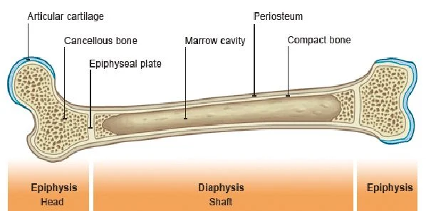
Radiotherapy of the long bones (RTLB) is meant to provide control of symptoms, destroy cancer cells in the treated area and prevent malignant disease-related fractures. Bone radiotherapy is also a useful means of halting tumour proliferation and then triggering osteoblastic activity with osteoproliferation. Bone recalcification after RTLB has been observed in 70 percent of cases, particularly in the fractionated group; recalcification commenced from the first month after RTLB and peaked at three months.
Nonetheless, RTLB does not entirely eliminate the risk of fracture, particularly in the three months immediately after radiotherapy. A pathologic fracture may occur during this time. While such fractures may be atraumatic, they can also considerably aggravate morbidity and mortality.
Numerous studies have explored the risk of fracture using radiographic images (standard x-rays) to determine predictive factors. Analysis of the dimensions of metastatic lesions, and especially any cortical involvement, on standard x-rays alone remains insufficiently predictive of the fracture risk. A three-dimensional CT study provides a more precise assessment of the risk of pathologic fracture, but is not always carried out when the pain is so great that radiotherapy is urgently required. CT scan-based virtual simulation is therefore a valuable tool for providing a precise analysis of tumour infiltration and osteolysis.
A study which has been published in Radiation Oncology aimed to use CT scan-based virtual simulation to assess the risk of fracture and identify predictive factors with a view to offering prophylactic fixation to those most at risk.
Materials and Method
The retrospective study was conducted in a single centre. Forty-seven (47) cancer patients were treated with RTLB (18 lung, 11 breast, 10 prostate and eight other cancers) between September 2010 and February 2012. The virtual simulation was carried using a 16-slice GE scanner with an 80 cm ring no more than three weeks prior to the start of radiotherapy. Each patient had been monitored for a minimum of four months in order to screen for post-RTLB fractures. Recalcification commenced from the first month after RTLB and peaked at three months; therefore, the fracture risk was considered low after the third month.
Two doctors (a radiotherapist and an oncologist) systematically analysed several parameters that are known from previous publications to be risk factors for pathologic fractures: the type and appearance of a metastasis, the mean dimensions of the lesion and the cortical involvement (CI) (craniocaudal, circumferential and thickness):
The statistical analysis was conducted using the Chi2 test and means comparison test. The performance characteristics of the craniocaudal and circumferential cortical lysis thresholds were analysed using the ROC curve. The value of p was considered significant when <0.05.
Results
The male gender ratio was 0.57 and the mean age was 62.8 (33–93) years. The average size of the metastatic lesions was 32 (8–87) x 2 (6–81) x 52 (7–408) mm, with cortical involvement (CI) in 66 percent of cases. The site was in the upper third of the bone in 92 percent of cases (28 femoral, 17 humeral and two tibial). Ten fractures occurred: two during RTLB, seven after one month and one after 6.6 months. The fractured lesions measured 48 (17–87) x 34 (12–66) x 76 (38–408) mm.
The predictive parameters for fracture were osteolytic (39 percent vs. 10 percent; p = 0.02) and permeative lesions (42 percent vs. 0 percent; p< 0.0005), a Mirels score ≥9 (42 percent vs. 0 percent; p< 0.0005), circumferential CI ≥30 percent (71 percent vs. 0 percent, p< 0.00001), CI ≥45 mm in height (67 percent vs. 0 percent, p< 0.00001) and CI in thickness =100 percent (40 percent vs. 0 percent; p = 0.0008). In the multivariate analysis, circumferential CI ≥30 percent was the only predictive parameter for fracture (p = 0.00035; OR = 62; CI 95 percent: 6.5-595).
Overall survival was 91 percent, 55 percent and 40 percent at one month, six months and one year, respectively.
Conclusions
This study analysed the risk of fractures following radiation for bone metastasis and attempts to determine which patient population would benefit from prophylactic surgery. Circumferential cortical involvement is easy to measure and should be systematic during CT scan-based virtual simulation prior to radiotherapy. Prophylactic primary fixation surgery should always be considered when the circumferential CI ≥30 percent.
The findings and data must be confirmed in a prospective study including a much larger series of patients, since it is important to precisely establish when prophylactic fixation is required to reduce morbidity and mortality. The development of an instrument that identifies patients who have a relatively high risk of developing such a fracture and therefore should be considered candidates for surgical stabilisation is helpful. This strategy could optimise the management of fragile metastatic patients.
Image Credit: BBC News
Nonetheless, RTLB does not entirely eliminate the risk of fracture, particularly in the three months immediately after radiotherapy. A pathologic fracture may occur during this time. While such fractures may be atraumatic, they can also considerably aggravate morbidity and mortality.
Numerous studies have explored the risk of fracture using radiographic images (standard x-rays) to determine predictive factors. Analysis of the dimensions of metastatic lesions, and especially any cortical involvement, on standard x-rays alone remains insufficiently predictive of the fracture risk. A three-dimensional CT study provides a more precise assessment of the risk of pathologic fracture, but is not always carried out when the pain is so great that radiotherapy is urgently required. CT scan-based virtual simulation is therefore a valuable tool for providing a precise analysis of tumour infiltration and osteolysis.
A study which has been published in Radiation Oncology aimed to use CT scan-based virtual simulation to assess the risk of fracture and identify predictive factors with a view to offering prophylactic fixation to those most at risk.
Materials and Method
The retrospective study was conducted in a single centre. Forty-seven (47) cancer patients were treated with RTLB (18 lung, 11 breast, 10 prostate and eight other cancers) between September 2010 and February 2012. The virtual simulation was carried using a 16-slice GE scanner with an 80 cm ring no more than three weeks prior to the start of radiotherapy. Each patient had been monitored for a minimum of four months in order to screen for post-RTLB fractures. Recalcification commenced from the first month after RTLB and peaked at three months; therefore, the fracture risk was considered low after the third month.
Two doctors (a radiotherapist and an oncologist) systematically analysed several parameters that are known from previous publications to be risk factors for pathologic fractures: the type and appearance of a metastasis, the mean dimensions of the lesion and the cortical involvement (CI) (craniocaudal, circumferential and thickness):
- Cortical thickness (mm) was the measurement of the thickness of the cortex considered to be normal in the most at-risk area.
- Craniocaudal cortical lysis (mm) was the measurement of the maximum CI height in the craniocaudal plane. The 30 mm threshold involvement was always recorded since this is the threshold predictive of pathological fracture according to several authors.
- Circumferential cortical lysis (mm) was the measurement of the diseased cortex perimeter in the most at-risk area. The threshold involvement of 50 percent was always recorded since this is the threshold predictive of pathologic fracture according to several authors.
- Cortical thickness lysis (mm) was the measurement of the maximum thickness of cortical lysis in the at-risk area. The percentage of cortical lysis thickness was always determined.
- The Mirels score takes into account anatomical location, extent of cortical lysis, appearance of the lesion and pain intensity and was calculated for each metastatic lesion. A Mirels score of nine or more was found to be predictive of fracture.
The statistical analysis was conducted using the Chi2 test and means comparison test. The performance characteristics of the craniocaudal and circumferential cortical lysis thresholds were analysed using the ROC curve. The value of p was considered significant when <0.05.
Results
The male gender ratio was 0.57 and the mean age was 62.8 (33–93) years. The average size of the metastatic lesions was 32 (8–87) x 2 (6–81) x 52 (7–408) mm, with cortical involvement (CI) in 66 percent of cases. The site was in the upper third of the bone in 92 percent of cases (28 femoral, 17 humeral and two tibial). Ten fractures occurred: two during RTLB, seven after one month and one after 6.6 months. The fractured lesions measured 48 (17–87) x 34 (12–66) x 76 (38–408) mm.
The predictive parameters for fracture were osteolytic (39 percent vs. 10 percent; p = 0.02) and permeative lesions (42 percent vs. 0 percent; p< 0.0005), a Mirels score ≥9 (42 percent vs. 0 percent; p< 0.0005), circumferential CI ≥30 percent (71 percent vs. 0 percent, p< 0.00001), CI ≥45 mm in height (67 percent vs. 0 percent, p< 0.00001) and CI in thickness =100 percent (40 percent vs. 0 percent; p = 0.0008). In the multivariate analysis, circumferential CI ≥30 percent was the only predictive parameter for fracture (p = 0.00035; OR = 62; CI 95 percent: 6.5-595).
Overall survival was 91 percent, 55 percent and 40 percent at one month, six months and one year, respectively.
Conclusions
This study analysed the risk of fractures following radiation for bone metastasis and attempts to determine which patient population would benefit from prophylactic surgery. Circumferential cortical involvement is easy to measure and should be systematic during CT scan-based virtual simulation prior to radiotherapy. Prophylactic primary fixation surgery should always be considered when the circumferential CI ≥30 percent.
The findings and data must be confirmed in a prospective study including a much larger series of patients, since it is important to precisely establish when prophylactic fixation is required to reduce morbidity and mortality. The development of an instrument that identifies patients who have a relatively high risk of developing such a fracture and therefore should be considered candidates for surgical stabilisation is helpful. This strategy could optimise the management of fragile metastatic patients.
Image Credit: BBC News
References:
Tatar Z, Soubrier M, Dillies A F, Verrelle P, Boisgard S, Lapeyre M
(2014) Assessment of the risk factors for impending fractures following
radiotherapy for long bone metastases using CT scan-based virtual
simulation: a retrospective study. Radiat Oncol. 2014; 9(1): 227. PMCID:
PMC4205287
Latest Articles
Radiotherapy, Fractures, metastases, bone, recalcification
Radiotherapy of the long bones (RTLB) is meant to provide control of symptoms, destroy cancer cells in the treated area and prevent malignant disease-relat...


