


HealthManagement, Volume 12 - Issue 5, 2012
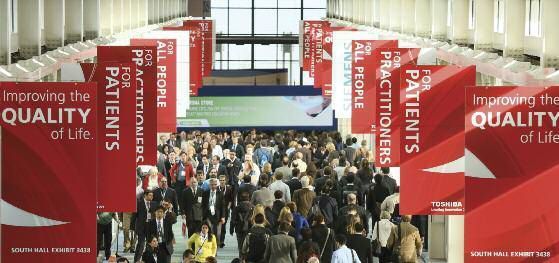
More than 53,000 delegates descended on Chicago in November for the 98th Radiological Society of North America Annual Meeting 2012. As well as scientific research, the future of radiology was a common theme, with new applications of technology, clinical research and the need for radiologists to become more visible all covered.

Putting Patients First: Rhetoric or Responsibility?
“Patients first” was the theme for this year’s Annual Meeting. RSNA President, George Bissett set the tone in his address, noting that recent developments in US healthcare gave even more compelling reasons for radiologists to demonstrate their patient-centredness. In the United States there are immense changes coming in an increasingly consumer driven and value-based healthcare environment, Bissett noted. If radiologists are invisible to patients, then they are vulnerable.
Said Bissett, “We have become focused on conveying information to the clinician for diagnostic purposes. We have developed fabulous technical capacities, instantly sending images all over the globe. In all of this it is easy to forget that a human being, with emotions and fears, is attached to the body parts we are studying. Patients have tremendous respect for our technology. However they are frustrated by things that many of us don’t have to deal with when we are studying the images – long waiting times, lack of information about procedures.
He urged his audience to walk a mile in their patients’ shoes and “step out from behind the curtain and put a face to the radiologist.” He himself had spent time in his own facility’s waiting room and recommended the audience to do the same, to talk to waiting patients, to look for ways to introduce themselves to patients. Eighty to ninety percent of radiologists never meet their patients, he noted. Other ways to become more visible are to provide information materials or add features to reports to help patients better understand their results.
Bissett strongly argued that the future for radiologists depends on their capacity to develop a new kind of shared ownership of their patients’ needs and expectations along with primary care and specialty colleagues. It was time, he said, to stop referring to ‘the patients’ and instead talk about ‘our patients.’
RSNA launched its Radiology Cares campaign at the meeting. The campaign is designed to help radiology professionals become more comfortable interacting directly with their patients, and to help patients become more comfortable with their radiology experiences. Radiologists are invited to take the Radiology Cares pledge. RSNA released a hard-hitting video series to illustrate what might happen if radiology did not become more patient-centred. The tenets of the campaign are that radiology professionals:
- Care for and about our patients.
- Treat every patient as we would a neighbour, friend or family member.
- See the patient behind the image.
- Align radiologic practice to serve our patients’ best interest.
In the opening session panel on putting patients first, two lung cancer survivors, Sheila Ross, an advocate for the Lung Cancer Alliance and Dr. Karen Arscott spoke eloquently of their experiences with radiology. Both made the point that they never met their radiologist, but met their radiation oncologist countless times. Ross urged radiologists to help get guidelines on lung cancer screening.She carries a small ball with her to show the size of what a chest X-ray can miss, but a CT scanner can pick up. Dr. Arscott recalled he her diagnosis with lung cancer,when she re ceived her diagnosis by post – “nobody called, nobody ever told me I had cancer.”
Radiology in Facial Transplantation
Professor Bohdan Pomahac presented on the remarkable facial transplantations performed by his team at Brigham and Women’s Hospital, work which relied on the highly expert radiology provided by his colleagues. He calls these highly advanced techniques facial restoration. Professor Pomahac told his audience, “We need your help to guide us to see what we can do, how far we can push and hopefully in the future, even intraoperatively guide where we can go and what can be done."
Radiology Informatics
When Professor Paul Chang’s father retired from radiology, Chang was surprised when his father accused him of ‘killing radiology’. While Chang junior thought his father alluded to laziness, he was in fact looking back to the time before PACS when medicine and surgery rounds started in radiology in the morning, when radiologists were the ‘doctor’s doctor’.
Professor Chang presented one of the Eugene P Prendergrass New Horizons Lectures.There is no going back, Chang acknowledged, even though digitisation has facilitated commoditisation and outsourcing. As radiology moves towards the third generation of PACS the emphasis is still on images, but with little discussion on the value added by radiologists. “Rarely do we interact”, Chang said, “It is easy to be commoditised and devalued when we are not seen.”
Chang suggested that the opportunity is there to be valued as radiologists managing the role of imaging in a capitated aligned system.Radiologists have to add value by optimising quality, safety and efficiency. IT is the way to do this – more capable and agile solutions are needed to provide measurable value in patient care. Intelligent business analytics can assist colleagues upstream and downstream and connect with health consumers. IT can enable electronic protocolling; the modality can also be treated as an IT device. Intelligent agents can use natural language processing to extract information from the electronic medical record (EMR).
“Radiology will not return to the personal contact of the pre-PACS era, but IT can enable radiologists to virtually collaborate with colleagues and patients”, Chang said.” He suggested radiologists view the radiology report as a portal. For example, the information in there can be hyperlinked to other systems, and presented in graphical or text form, e.g. contrast or radiation dose. Chang closed by saying the challenge is to re-engineer ourselves as radiologists. "The technology is easy", he acknowledged. "Changing human behaviour and legacy workflow is much harder.
" Chang and colleagues from the University of Chicago presented on their Annotation and Image Markup (AIM)-based lesion tracking tool. The tool is integrated into PACS and has shown significant improvements in oncologic lesion measurement efficiency and error reduction.
Professor Keith Dreyer of Harvard University and Massachusetts
General Hospital presented the second Eugene P Prendergrass New Horizons
Lecture on the future of imaging informatics. Dreyer explored changes in
healthcare and the informatics innovation necessary to remain relevant and
effective in the rapidly evolving healthcare system. He argued that previous
payment models have determined business models for radiology in the United States. Fee-for-service has incentivised volume,
while being neutral on value.Business models have determined innovation,
maximising productivity and value while reducing the cost of doing business.
Innovation is different depending on whether the incentives are for volume
or outcome. In the context of patients first, why not extend functionality to
your patients? The meaningful use programme in the U.S. provides incentives for radiologists to participate and promotes the use of certified
electronic health record technology (CEHRT) to improve the safety, quality and
cost of health care. Dreyer noted that there are opportunities for innovation
in the current healthcare climate, in access, communication and utilisation.
Currently imaging information is too compartmentalised, and he looked forward
to RIS and PACS being converged into the electronic health record. He
anticipated a future of structured, multimedia, interactive communication.
Structured reporting with clinical decision support will enable actionable
findings and actionable recommendations. There are limited ways for radiologists to participate in management and ordering of
examinations. However, the EHR linked to the American College of Radiology
(ACR) Approriateness Criteria enables communication between the ordering
physician and the radiologist. For the future, he said, image sharing, structured
reporting in the EHR and personal health records will increase quality, and
most importantly, the radiologist’s presence and importance.

The Story Behind the Image
Professor Richard Gunderman from Indiana University talked eloquently about the need to delve deeply to find the story behind the image. He recalled reading the CT scan of an QJ year old man with dementia and a history of falls. It turned out that his patient was the Nobel Prize winner, Charlie Huggins.
Said Gunderman, “Radiology like all human endeavours makes it possible to get the technical aspects right. We can be extraordinarily precise and utterly inaccurate. We can get the little things right and completely neglect what matters most. We can develop a perfect industrial model of clinical radiological practice and end up laying waste to the humanity of radiologists and more importantly the patients we serve.”
Gunderman urged radiologists to focus more time and attention on the human excellence of radiologists, and asked, “When was the last time you heard a great story about what it means to be a radiologist?”
He acknowledged that although radiologists do not have time to delve deeply into each image we should treat each as possessed of the degree of preciousness that he found in the head CT scan of Charlie Huggins.
He concluded, “We cannot put patients first unless we first know our patients, and that means being acutely attuned to the story that lies behind the image.”
The Visible Radiologist
The need to become more visible was underlined by recent research in Indiana, which showed that only 53.5% of patients surveyed after undergoing a CT scan knew that a radiologist was a doctor. Study author, Dr. Peter Miller, of Indiana University School of Medicine, said, "We need to better understand what patients want to know about radiologists in order to improve service and patient care. In my experience, people who've had the opportunity to interact with radiologists appreciated the chance to talk with them and get their thoughts on the imaging results."
Medicolegal Issues
Dr. Leonard Berlin gave the keynote session on “To disclose or not to disclose.” Delivered entirely in rhyming couplets, his presentation poetically urged radiologists to disclose errors completely, regardless of type, severity, cause or frequency. Healthcare has moved from the era of 'doctor knows best' to an era of patient individualism, participation and shared decision-making where doctors should inform but not influence. Concern about liability should not diminish this duty.Doctors should be concerned and sympathetic following and error and be apologetic if it is their fault. "Avoid the appearance of wrongdoing; would that stop the patient from suing?" he said. In closing, he said, “Remember, an apology is not an elegy.”
A mock jury trial mediated by Dr. Berlin presented a fictitious case of a radiologist sued by the widower of a woman who had died from breast cancer after five CT exams and a CT coronary angiogram.
Did the jury decide that the radiologist was negligent for not alerting the referring physician and/or the patient that radiation exposure from CT scans may cause breast cancer? The radiologist’s defence was that the evidence for a link between diagnostic radiation exposure and cancer is unproven. The trial ended with a split jury.
And the take-home message? Professor Rebecca Smith-Bindman, who
testified for the plaintiff, recommended that radiologists should not only
ensure that all exams are performed at the lowest possible dose needed to
achieve a diagnostic quality image, but they also should ensure that there are
processes to identify questionable exams and communicate the possible risks to
the referring doctor.
Imaging of Inpatients
Dr Shima Aran presented a study comparing wireless direct radiography (DR) and computed radiography for portable chest radiography (CR) in the intensive care unit.The researchers concluded that portable chest radiography using wireless DR in the ICU setting provides similar or superior information on clinically significant findings while retaining the image quality of CR. Visualisation of some anatomic landmarks and tubes and lines was superior with DRw compared to CR. The wireless system enabled faster turnaround time and smoother workflow, and with no wire, reduced risk of breaking the sterile field.
Dr Arielle Lutterman and colleagues at Emory School of Medicine looked at cumulative radiation exposure of hospitalised patients due to imaging and image-guided procedures. Many patients are unaware of these risks, and their cumulative exposures are not being tracked. Their study looked at the records of 200 inpatients in two urban university hospitals.
They found that hospitalised patients are experiencing high levels of radiation exposure during a single hospitalisation. KH% of hospitalised patients underwent CT scanning, and the majority (82%) of inpatient radiation exposure was attributable to CT scans. The mean dose estimate per patient for one hospitalisation was 14.76 mSv with 82% of the radiation exposure due to CT examinations.
In their study eleven patients (5.5%) received 5O mSv, two received >100 mSv. Fifty one patients received 20-49 mSv. They recommend that radiologists should be vigilant in terms of monitoring CT imaging protocols so that when a CT is ordered, the resultant radiation exposure will be as low as is reasonably achievable. Consideration should be given to alternative forms of imaging such as magnetic resonance imaging when possible and based on a given institution's expertise.
Breast Cancer Risk from CT and Nuclear Imaging
Researchers who reviewed the records of approximately 250,000 women enrolled in an integrated healthcare delivery system between 2000 and 2010 found that increased CT utilisation could result in an increase in the risk of breast cancer for certain women, including younger patients and those who received repeat exams.
"Young women receiving several chest and or cardiac CTs had the greatest increased risk of developing breast cancer at approximately HR percent," said Diana Miglioretti, Ph.D., study coauthor and senior investigator at the Group Health Research Institute. "A 15-year-old girl with no risk factors for breast cancer would double her 10-year risk of developing breast cancer at 25.
"To lower imaging-related risk of developing breast cancer, senior author Professor Rebecca Smith-Bindman said imaging providers should analyse the radiation doses associated with each exam, reduce the use of multi-phase protocols and employ dose-reduction software wherever possible to minimise exposures.
"If imaging is truly indicated, then the risk of developing cancer is small and should not dissuade women from getting the test they need," she said. "On the other hand, a lot of patients are undergoing repeat chest and cardiac CT, many of which aren't necessary. Women, and particularly young women, should understand there is a small but real potential risk of breast cancer associated with cardiac and chest CT, and the risk increases with the number of scans.”
"The researchers found a wider than expected variation in
dose for exams. The dose for certain paediatric and adult exams was more
similar than expected. For nuclear medicine however, there was an appropriate
lower dose for paediatric patients compared to adults.
Industry Show
Agfa Healthcare
Carestream
Carestream’s MyVue software offers patients access to their own images. A trial of the technology in Houston with 2000 patients saved cUS$7 per image compared to CDs. Patient satisfaction was also very high.
GE Healthcare
GE demonstrated its Silent Scan MRI (ONRk pending) with a live link-up to its testing facility.The decibel level of conventional MRI scanners can be more than NRR decibels. The noise from the new scanner, which uses a new pulse sequence, is comparable to having an inkjet printer running in the room next door.
GE launched DoseWatch N.H which has a size specific dose estimate, and compares scan time to conventional scan times. It is integrated with radiology dictation software.
Universal Viewer is an advanced workstation embedded in the PACS. It is designed to aid informed decision making by providing historical data. It includes unified web tools for productivity, enables cross enterprise collaboration and consolidates patient history.

Philips
Philips presented several new products, including:
- The latest version of its IntelliSpace Portal, an advanced visualisation solution for the analysis and interpretation of medical images designed to simplify the way radiologists work with the vast amounts of imaging data sets.
- iPatient, an advanced platform for its family of CT and PET/CT scanners. iPatient allows for easy and efficient communication between the CT system and the injector in order to deliver appropriate contrast dose and consistent image quality.
- The launch of a cost-effective alternative to provide digital broadband MRI. The revolutionary dStream broadband technology, which Philips introduced with its Ingenia MR systems, provides enhanced image quality, improved workflow, easier coil handling and better patient comfort.
- With the next generation Ingenia MR-OR solution for intraoperative neurosurgery, Philips is further expanding its MR offering in the interventional MRI area. An MR-OR suite for intraoperative MRI adds value to neurosurgical facilities, supporting resection procedures that can save precious time for both surgeon and patient.
- The next generation of MicroDose with Single-Shot Spectral Imaging (SI) together with the first clinical application – Spectral Breast Density Measurement. The new Philips MicroDose SI, a full-field digital mammography system, brings the potential of non-invasive spectral imaging to clinical practice without exposing women to additional examinations or X-ray radiation.
Siemens
Interest was high in Siemen’s world-first wireless ultrasound, the ACUSON Freestyle™.With a range of up to three metres, the ability to control the device via an ergonomic interface enables remote control of scanning parameters from within the sterile field. The system’s innovations include acoustics, system architecture, radio design, miniaturisation and image processing.
“Siemens Healthcare is the first company to introduce an ultrasound system that enables physicians to work with cable-free transducers,” said Jeffrey Bundy, CEO of the Siemens Healthcare Ultrasound business unit.“The ACUSON Freestyle system facilitates the use of advanced ultrasound technology into clinical fields requiring a sterile environment, such as interventional radiology, anaesthesiology, critical care, cath lab, or emergency care.”
The ACUSON Freestyle system employs advanced synthetic aperture imaging technology, an integration of proprietary hardware and software that was specifically developed for the wireless signal transmission of full-resolution digital image data at very high data rates. Focusing on each pixel in the image, this method produces excellent image quality throughout the field of view. This design reduces the transducer’s power requirements, increasing battery life. The battery can be sterilised. Wireless real-time ultrasound data transmission is further enabled through the proprietary development of a novel ultra-wideband radio technology, which, operating at a high frequency of 7.8 Gigahertz, is not susceptible to interference with other electronic equipment. The system includes exam presets, automated features including focus and it enables image save and clip save. It has 16GB storage, DICOM capabilities and wifi built in.
Three wireless transducers are available for the ACUSON Freestyle system, covering a range of general imaging, vascular, and high-frequency applications such as musculoskeletal and nerve imaging. The ACUSON Freestyle system has a MQ-centimeter, highresolution LED display.
The ACUSON Freestyle is 510k cleared and is expected to ship in
summer 2013. Siemens’ works-in-progress imaging systems include:
- MAGNETOM Prisma, a 3 Tesla MRI scanner designed to combine high gradient strength and fast gradient slew rates with a significantly higher signal-to-noise ratio to tackle demanding clinical and research challenges. It is a Diffusion Spectrum Imaging (DSI) application designed to resolve fine anatomical details of the brain.
- Cios Alpha, a mobile C-arm system designed with greater power output and a larger field of view in the operating room (OR) than conventional C-arms. It is intended to create high-quality images in the field of vascular
- MAGNETOM Essenza with Siemens’ MRI workflow solution Dot (Day optimizing throughput), which helps enhance productivity in MR scanning.
- MAGNETOM Trio, Verio, and Avanto upgrades with Siemens’ MR workflow solution Dot (Day optimizing throughput), which helps enhance productivity in MR scanning, as well as Tim (Total imaging matrix) 4G – the latest generation of Siemens’ integrated coil technology.
- syngo.via WebViewer VA11, which is intended to perform diagnostic reading directly on the iPad as well as provide access to images from computed and digital radiography (CR and DR), positron emission tomography (PET), and PET/ computed tomography (CT) devices.
RSNA 2013 will be back in Chicago from December 1-6, with the theme of “The Power of Partnership.”