The rising incidence of renal cell carcinoma in Western populations, coupled with advancements in radiological imaging, has led to increased detection of renal tumours, particularly small renal masses (SRMs). While this improved detection offers potential for early intervention, it also raises concerns about overdiagnosis and overtreatment. Accurate segmentation of renal tumours is crucial for better diagnosis, treatment planning, and surgical decision-making. Recent developments in automatic renal tumour segmentation using neural networks offer promising solutions to these challenges, enabling more precise and efficient care.
The Challenge of Detecting Small Renal Tumors
The widespread use of cross-sectional radiological imaging has significantly improved the detection of renal tumours. However, small tumours, particularly those less than 4 cm in diameter (T1a stage), pose a diagnostic challenge. These tumours are often difficult to characterise radiologically due to their subtle imaging features. As a result, patients in regions with high scanning rates face increased risks of undergoing unnecessary partial or total nephrectomy or renal ablation, which may indicate overdiagnosis and subsequent overtreatment. Therefore, more accurate methods for identifying and assessing these small renal tumours are needed to ensure that only those requiring intervention are treated.
\
The Role of Renal Tumor Segmentation in Treatment Planning
Renal tumour segmentation is a vital step in evaluating the size and location of tumours, which directly impacts treatment planning. Accurate segmentation aids in volumetry and surgical planning, including determining the complexity of renal tumours, as seen in tools like the R.E.N.A.L. nephrectomy score. This score helps surgeons decide on the most appropriate surgical approach and follow-up care. Moreover, precise tumour segmentation is essential for planning minimally invasive procedures such as thermal ablation, where the accurate placement of ablation probes and margins is critical. Therefore, improving segmentation accuracy directly contributes to better patient outcomes.
Advancements in Automatic Renal Tumor Segmentation
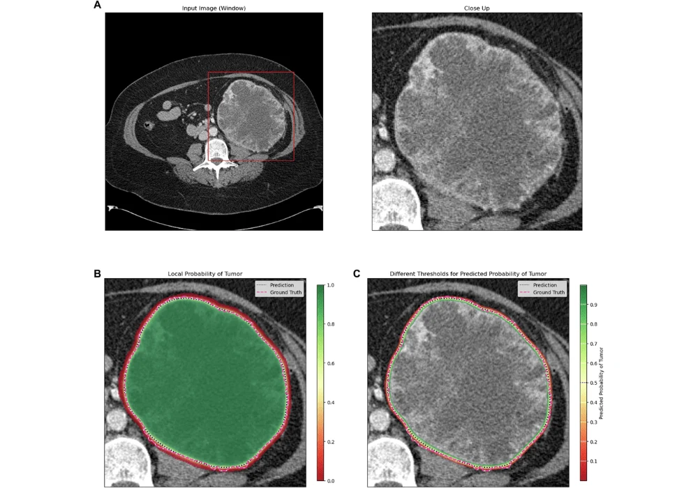
Manual segmentation of renal tumours, though well-established, is labour-intensive and prone to variability among radiologists. Researchers have developed automatic renal tumour segmentation algorithms using neural networks to overcome these limitations. These algorithms, such as the Ensemble Neural Network (ENN) model, have shown great promise in automating and refining the segmentation process. In a recent study, ENN models were trained on a large dataset of contrast-enhanced CT images from patients with renal tumours. The results demonstrated that the ENN could accurately segment renal tumours with high confidence, as evidenced by a median DICE score of 0.84. This performance was consistent across different datasets, highlighting the model's robustness and potential for widespread clinical application.
Conclusion
The integration of advanced neural networks into renal tumour segmentation represents a significant leap forward in detecting and treating renal tumours. By automating the segmentation process, these models reduce the burden on radiologists and increase the accuracy and consistency of tumour assessments. Moreover, the ability of these models to provide visual feedback and confidence measures enhances their clinical utility, aiding in surgical planning and reducing the risks associated with overdiagnosis and overtreatment. As these technologies continue to evolve, they hold the potential to improve patient outcomes and streamline the management of renal tumours. Further studies are needed to refine these models and evaluate their impact in clinical settings. Still, the future of renal tumour treatment looks increasingly promising with the aid of artificial intelligence.
Source & Image Credit: European Radiology

