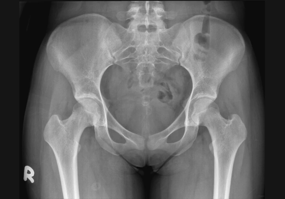
Pelvic organ prolapse (POP) is common in later life and its impact is set to grow. Estimates suggest that around one in ten women over 50 are affected, with numbers expected to rise by midcentury. Known risk factors such as parity, vaginal delivery, levator ani defects, urogenital hiatus size and neonatal birth weight are usually recognised after childbirth, while symptoms may not appear until years later. Earlier risk insight could help women and clinicians plan care and prevention. Using magnetic resonance imaging (MRI) and shape analysis, researchers compared pelvic floor soft tissue and the bony pelvis in parous women with POP, parous controls and nulliparous women to explore whether pelvic shape itself is linked to POP and whether the same signal is visible in younger adults.
Imaging Highlights Distinct Patterns
The main dataset included women around 50 years of age split across three groups: parous women with POP, parous controls and nulliparae. Three dimensional landmarks were placed on MRI scans to capture both the soft tissues that support the pelvic floor and the bones that form the pelvic canal. Configurations were standardised to focus on shape rather than size or position, and key patterns of variation were then identified.
Must Read: Recognising Incidental Findings in Female Pelvic MRI
When soft tissue and bone landmarks were assessed together, the three groups separated along the main shape patterns. POP cases tended to have a wider urogenital hiatus, extending partly below the ischiopubic outline. Nulliparous women showed a narrower hiatus positioned higher above the ischial bones. Parous controls sat between these two configurations. Within the combined analysis, the overall form of the bony pelvis also differed, with POP cases showing a pelvis that was shallower from front to back, wider side to side and shorter from top to bottom than the other groups.
Soft tissue differences alone were present but less decisive for risk assessment. A larger urogenital hiatus and lower muscle position were seen with POP, yet soft tissue changes were also observed between parous controls and nulliparae, suggesting sensitivity to childbirth and possible change over time. In contrast, bony shape varied in a way that aligned consistently with POP status among parous women and also showed a similar pattern in younger adults.
Bone Structure Separates Groups
Focusing only on bony landmarks strengthened the distinction between parous women with POP and parous controls. Along the main shape axis, the groups showed almost complete separation with strong statistical support. POP cases were characterised by a pelvis that was wider mediolaterally, shallower anteroposteriorly and shorter craniocaudally, with a wide subpubic angle. Parous controls had a taller pelvis with a relatively expanded anteroposterior dimension, producing a round to oval canal and a narrower subpubic angle. Nulliparous women spanned the range between cases and controls yet still differed from cases on the same axis.
The size of the difference across groups was large. Correlations between the principal shape signal and basic body measures such as height or body mass index were weak, indicating that the observed pattern was not simply a consequence of overall body size. To examine whether the same signal can be seen earlier in life, a small separate dataset of parous women in their early thirties was projected into the same shape space. The two younger controls mapped within the control region, and one younger POP case aligned with the case region while the other lay close to the boundary. This suggests that the pelvic shape pattern associated with POP in midlife can be detected in younger adults, although wider sampling is needed.
These findings build on the shift from manual pelvimetry to imaging based assessment. While traditional pelvimetry informed delivery planning, modern imaging offers a more detailed and less invasive way to quantify pelvic form. The present analysis indicates that bone shape carries information relevant to POP risk beyond the context of labour alone.
Towards Practical Assessment
To translate three-dimensional shape patterns into clinic friendly measures, the researchers derived a set of linear and angular variables from pelvic bone landmarks. These included ratios of width to depth and height, specific distances across the pelvic canal and measurements that reflect the spacing around the ischial spines and acetabular walls. Discriminant analyses compared the ability of these measures to separate groups against established characteristics such as body mass index, parity and levator ani injury.
Variables with the largest weights included the biacetabular width to depth ratio, inferior symphysis to coccyx depth, bispinal width, bispinal width to pelvic canal height ratio, bispinal width to depth ratio and the distance between acetabular walls. In this set, the spread of values did not overlap between cases and controls for bispinal width, the ratio of the midtuberosities to superior pubis height and body mass index, indicating potential utility for distinguishing groups. While soft tissue features like urogenital hiatus size were associated with POP, their variability across parity groups makes them less suited as standalone markers for risk stratification.
Importantly, the shape-based approach does not rely on changes that may follow childbirth or disease progression. By focusing on stable bony architecture, it may help identify individuals with a predisposition to POP irrespective of soft tissue status. However, the work points to the need for larger and more diverse datasets to define thresholds, test generalisability and understand performance across different populations and life stages.
MRI-based shape analysis identified a distinct bony pelvic configuration in parous women with POP compared with parous controls of similar age. Cases typically showed a pelvis that was wider side to side, shallower front to back and shorter top to bottom, with clear separation from controls and evidence of the same signal in a small group of younger adults. Clinical translations of these three-dimensional patterns into simple linear and angular measurements showed promise for discriminating cases from controls and could support counselling after delivery. Further validation is required to determine how these measurements might be used to inform risk discussions and care planning while avoiding reliance on soft tissue markers that may shift over time.
Source: Ultrasound in Obstetrics & Gynecology
Image Credit: iStock


