


Renal cell carcinoma (RCC) stands as the most lethal urological malignancy, particularly due to its poor prognosis in metastatic stages, with a reported 5-year cancer-specific survival rate ranging from 0 to 32% . Early detection and precise characterisation of renal tumours significantly enhance clinical management and patient survival. However, current diagnostic imaging methods fail to accurately differentiate between tumour grades and subtypes. This article explores the potential of novel T2 mapping methods in magnetic resonance imaging (MRI) to noninvasively characterise renal tumour subtypes and grades.
Challenges in Current Diagnostic Methods
Renal mass biopsy (RMB) is often used to diagnose RCC but has limitations, including invasiveness and a nondiagnostic rate of up to 14%. Additionally, RMB is susceptible to sampling errors due to tumour heterogeneity, particularly in clear cell renal cell carcinoma (ccRCC), potentially leading to inaccurate grading or unnecessary surgeries for benign lesions. Although traditional imaging methods can address some limitations of RMB, they primarily assess tumour size, lacking the ability to offer detailed microstructural and biological insights.
T2-weighted MRI images, integral to tumour evaluation, provide qualitative or semi-quantitative data on T2 relaxation times, which vary with molecular motion within tissues. T2 mapping offers robust quantitative assessments, allowing for direct comparisons within and between patients, and is promising in distinguishing high-grade from low-grade ccRCCs.
The TEMPURA Method: A Novel Approach
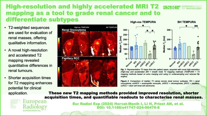
A novel technique, T2 mapping using the sequence Echo Merging Plus k-t Undersampling with Reduced refocusing flip Angles (TEMPURA), has been developed. TEMPURA enhances spatial and temporal resolution, producing high-resolution T2 maps within a single breath-hold, making it suitable for routine clinical use. TEMPURA’s high-resolution and breath-hold capabilities offer detailed microstructural visualisation and quantitative measures of intratumoral heterogeneity, which are crucial for accurate tumour characterisation.
The TEMPURA method was tested on patients recruited from the Uro-oncology Clinic at Addenbrooke’s Hospital, Cambridge, between January 2022 and January 2023. This study aimed to quantify T2 metrics across different renal tumour subtypes and grades and to evaluate the potential of TEMPURA in improving diagnostic accuracy.
Quantitative Results from T2 Mapping
Quantitative T2 mapping showed promising results in differentiating renal tumour subtypes and grades. For instance, the lowest median T2 values were found in papillary RCC (pRCC), significantly differing from other subtypes like renal oncocytoma (RO), chromophobe RCC (chRCC), and ccRCC. This differentiation is crucial as it can guide appropriate treatment strategies. Additionally, high-resolution TEMPURA provided greater microstructural detail compared to standard T2-weighted imaging, enhancing the visualisation of intratumoral heterogeneity.
For ccRCC, T2 mapping revealed a trend of decreasing median T2 values from lower to higher WHO/ISUP grades, indicating its potential to noninvasively predict tumour aggressiveness. Moreover, TEMPURA sequences showed good agreement in quantitative readouts, suggesting reliability in clinical settings.
The novel T2 mapping techniques, particularly the TEMPURA method, show significant promise in the noninvasive characterisation of renal tumours. By providing detailed insights into tumour microstructure and heterogeneity, these methods could improve the accuracy of subtype differentiation and grade prediction, ultimately enhancing patient management and treatment outcomes. Future studies should focus on larger patient cohorts and comparative analyses with other advanced MRI techniques to validate these findings and establish the clinical utility of TEMPURA in routine practice.
Source: European Radiology Experimental
Image Credit: iStock