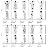
A groundbreaking study has introduced a pioneering method to predict lymph node metastasis (LNM) in patients with non-functional pancreatic neuroendocrine tumours (NF-PanNETs), aiding in tailored treatment strategies and enhancing patient outcomes. Recently published in eClinicalMedicine, the research team from the University of Tsukuba harnessed advanced imaging technology to develop and validate a novel predictive model with a 89% success rate in predicting LNM.
Addressing Limitations in Current Guidelines for surgery
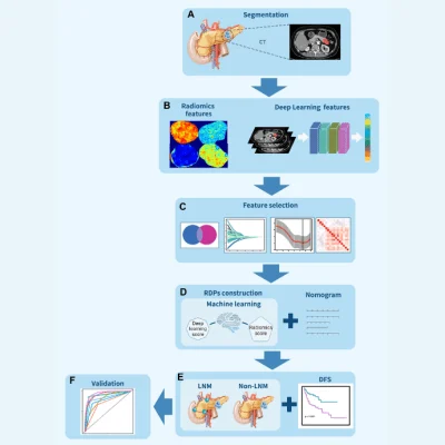
Non-functional pancreatic neuroendocrine tumours (NF-PanNETs) present unique challenges due to their varied biological behaviours and lack of clear guidelines for surgical intervention. Current clinical guidelines lack consensus on the threshold for lymph node dissection, particularly for tumours ≤2 cm. The study's findings fill this gap by offering a precise tool for assessing LNM risk, thereby ensuring tailored treatment strategies and avoiding unnecessary interventions. The study aimed to address these challenges by leveraging contrast-enhanced CT images and sophisticated machine-learning techniques. The research involved a comprehensive analysis of data from 320 patients across two medical centres, covering a period from January 2010 to March 2022. The team developed Radiomics Deep Learning Signatures (RDPs) based on ten machine-learning techniques, integrating these signatures with clinicopathological factors into a predictive nomogram for clinical use.
CT-based radiomics in support of surgical decision-making
The results were striking, with RDPs demonstrating exceptional performance in predicting LNM and disease-free survival (DFS) across both medical centres. Notably, the combination nomogram exhibited even higher predictive capability for LNM. Importantly, the model maintained effectiveness across different tumour sizes, providing valuable insights for surgical decision-making. The implications of this study are profound for clinical practice. By accurately predicting LNM preoperatively, the model offers valuable guidance for surgical planning, particularly regarding the extent of lymph node dissection. For tumours ≤2 cm, a low RDPs indicates a reduced likelihood of LNM, allowing for less invasive procedures or confident follow-up. Conversely, a higher RDPs suggests the necessity for standard oncologic resection. Similarly, for tumours >2 cm, the model aids in distinguishing between minimally invasive approaches and standard resection based on LNM risk.
Deep learning enables personalised medicine
While the study marks a significant advancement, it acknowledges certain limitations, including sample size constraints and the need for further validation in larger, multicentre studies. Additionally, integrating additional imaging modalities could enhance the model's robustness in clinical practice. The innovative predictive model presented in this study represents a paradigm shift in the management of NF-PanNETs. By harnessing cutting-edge imaging technology and machine learning, it offers clinicians a powerful tool for personalised treatment planning, ultimately improving patient outcomes and quality of life.
Source & Image Credit: eClinicalMedicine

