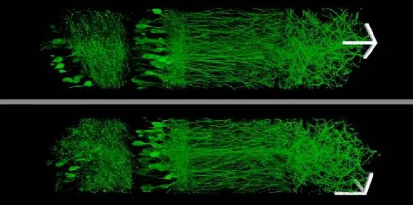
MIT researchers have developed a new technique that allows biological specimens to be physically magnified and then imaged at high resolution without needing to use very powerful — and often expensive — microscopes. This new method enlarges tissue samples by embedding them in an expandable polymer gel made of polyacrylate.
The expansion microscopy process or technique uses inexpensive, commercially available chemicals and microscopes commonly found in research labs. This innovation should give many more scientists access to "super-resolution" imaging, according to the MIT research team. (The latest generation of so-called "super-resolution" microscopes can see inside cells with resolution better than 250 nanometres.)
"Instead of acquiring a new microscope to take images with nanoscale resolution, you can take the images on a regular microscope. You physically make the sample bigger, rather than trying to magnify the rays of light that are emitted by the sample," says senior author Ed Boyden, an associate professor of biological engineering and brain and cognitive sciences at MIT.
MIT graduate students Paul Tillberg and Fei Chen are lead authors of the study, and their findings have been published in the online edition of Science.
How the Technique Works
Before enlarging the tissue, the MIT researchers first label the cell components or proteins that they want to examine, using an antibody that binds to the chosen targets. This antibody is linked to a fluorescent dye, as well as a chemical anchor that can attach the dye to the polyacrylate chain.
Once the tissue is labelled, the researchers add the precursor to the polyacrylate gel and heat it to form the gel. They then digest the proteins that hold the specimen together, allowing it to expand uniformly. The specimen is then washed in salt-free water to induce a 100-fold expansion in volume. While the proteins have been broken apart, the original location of each fluorescent label stays the same relative to the overall structure of the tissue because it is anchored to the polyacrylate gel.
"What you're left with is a three-dimensional, fluorescent cast of the original material. And the cast itself is swollen, unimpeded by the original biological structure," Tillberg explains.
Prof. Boyden et al. imaged this "cast" with commercially available confocal microscopes, commonly used for fluorescent imaging but usually limited to a resolution of hundreds of nanometres. With their enlarged samples, the MIT team achieved resolution down to 70 nanometres. "The expansion microscopy process ... should be compatible with many existing microscope designs and systems already in laboratories," Chen says.
Imaging Large Tissue Samples
The new technique enabled the MIT team to image a section of brain tissue 500 by 200 by 100 microns with a standard confocal microscope. Imaging such large samples would not be feasible with other super-resolution techniques, which require minutes to image a tissue slice only 1 micron thick and are limited in their ability to image large samples by optical scattering and other aberrations.
"The other methods currently have better resolution, but are harder to use, or slower," Tillberg points out. "The benefits of our method are the ease of use and, more importantly, compatibility with large volumes, which is challenging with existing technologies."
The new method could be very useful to scientists aiming to image brain cells and map how they connect to each other across large regions. "There are lots of biological questions where you have to understand a large structure," Prof. Boyden notes. "Especially for the brain, you have to be able to image a large volume of tissue, but also to see where all the nanoscale components are."
Prof. Boyden's team may be focused on the brain, although he mentions other possible applications for this technique including studying tumour metastasis and angiogenesis (growth of blood vessels to nourish a tumour), or visualising how immune cells attack specific organs during autoimmune disease.
Source: Massachusetts Institute of Technology
Image Credit: MIT
References:
Chen F, Tillberg PW, Boyden ES (204) Expansion microscopy. Science, Published online 15 January. DOI: 10.1126/science.1260088
Latest Articles
Brain, metastases, proteins, tumour, polymers, microscope
MIT researchers have developed a new technique that allows biological specimens to be physically magnified and then imaged at high resolution without needi...


