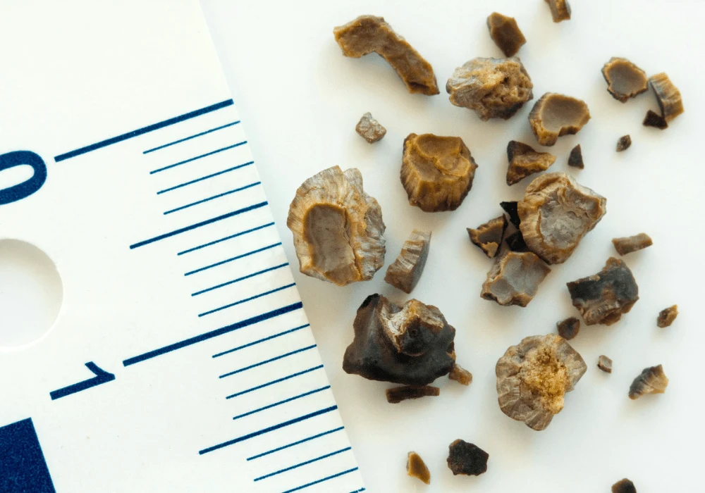
Kidney stones are a widespread medical condition, often resulting in intense pain and a significant healthcare burden. Accurate diagnosis is crucial for effective treatment, as the chemical composition of the stones plays a pivotal role in determining the appropriate therapeutic approach. While traditional imaging methods, such as conventional computed tomography (CT) and dual-energy CT (DECT), provide valuable diagnostic insights, they are limited in their ability to differentiate between stone subtypes with precision. Recent advancements in imaging technology, particularly photon-counting computed tomography (PCCT) combined with radiomics, have opened new frontiers for non-invasive, high-accuracy kidney stone classification. These technologies have the potential to revolutionise personalised treatment and preventive care strategies.
The Evolution of Imaging in Urolithiasis
Urolithiasis, characterised by the formation of stones within the urinary tract, has historically relied on non-contrast CT scans for diagnosis. These scans provide crucial information on the size and location of stones, facilitating treatment planning. However, the inability of conventional CT to determine the chemical composition of stones limits its usefulness in tailoring therapeutic approaches. DECT improved upon this by enabling the distinction between uric acid and non-uric acid stones, an advancement that informed decisions regarding pharmacological treatments and surgical interventions. Despite this, DECT exhibits reduced accuracy for smaller stones and at lower radiation doses, underscoring the need for further innovation.
Photon-counting CT has emerged as a groundbreaking technology, utilising advanced photon-counting detectors to overcome the limitations of traditional CT. By capturing individual X-ray photons and distinguishing between energy levels, PCCT enhances tissue contrast and reduces artefacts, significantly improving diagnostic clarity. This innovation offers superior imaging and enables multi-energy analyses, a critical capability for identifying the complex compositions of kidney stones. As a result, PCCT represents a significant leap forward in diagnosing and managing urolithiasis.
The Power of Radiomics in Kidney Stone Classification
Radiomics is a transformative field that extracts quantitative data from medical images, analysing patterns and features surpassing human perception limits. It involves the assessment of texture, shape and intensity at the pixel level, yielding biomarkers that provide detailed insights into the composition and characteristics of kidney stones. When paired with PCCT, radiomics have demonstrated remarkable potential in classifying various stone types with high accuracy.
The study outlined in the document highlights the integration of radiomics and PCCT for the differentiation of six kidney stone subtypes, achieving an impressive accuracy rate of 81% and an area under the curve (AUC) of 0.95. By employing random forest classifiers and monoenergetic reconstructions across multiple energy levels, the researchers were able to extract key radiomics features that are integral to stone classification. Parameters such as texture strength and wavelet-based variance emerged as significant contributors to distinguishing between stones composed of materials like uric acid, cystine and struvite. The combination of PCCT's spectral imaging capabilities and radiomics' quantitative analysis enables a nuanced understanding of stone characteristics, paving the way for tailored treatments and improved patient outcomes.
This radiomics-driven approach also reduces reliance on invasive procedures for stone analysis. Conventional methods often involve retrieving stones surgically for laboratory examination, an invasive process that carries risks for the patient. The ability to characterise stones non-invasively through PCCT imaging and radiomics could transform clinical workflows, enhancing diagnostic efficiency and patient comfort.
Clinical Implications and Future Directions
The clinical applications of combining PCCT with radiomics are profound and far-reaching. Accurate differentiation of kidney stone subtypes enables clinicians to adopt personalised treatment strategies. For instance, uric acid stones, which can often be dissolved using alkalising agents, require a different approach from other types, such as calcium oxalate or struvite stones. Misclassification could result in inappropriate therapies, potentially leading to complications or stone recurrence. With PCCT and radiomics, treatment decisions can be more precise, minimising recurrence rates and improving patient outcomes.
Beyond its diagnostic capabilities, PCCT also addresses concerns about radiation exposure, a key issue for patients requiring frequent imaging. By improving image quality at lower doses, PCCT ensures safer diagnostic procedures without compromising accuracy. This advantage is particularly relevant in the management of chronic urolithiasis, where repeated imaging is often necessary for monitoring and follow-up.
Looking to the future, integrating artificial intelligence (AI) and deep learning with radiomics offers exciting possibilities for further enhancing diagnostic workflows. AI algorithms can analyse vast datasets more efficiently, identifying subtle patterns and correlations that might elude conventional methods. Moreover, deep learning-based image enhancement techniques have already shown promise in improving image quality and reducing noise in low-dose CT scans. Incorporating these advancements into PCCT-based diagnostics could further refine the classification of kidney stones while maintaining minimal radiation exposure.
In addition, the use of PCCT and radiomics is not confined to kidney stone analysis. These technologies have potential applications in oncology, where they could aid in distinguishing tumour subtypes or predicting genetic mutations, effectively serving as virtual biopsies. The versatility of PCCT positions it as a valuable tool across multiple medical imaging domains.
The advent of photon-counting CT and its integration with radiomics heralds a new era in diagnosing and managing kidney stones. Combining advanced imaging technology with robust data analysis offers precise, non-invasive differentiation of stone subtypes, enabling clinicians to tailor treatments to individual patients. The potential benefits extend beyond immediate diagnosis, offering opportunities for personalised prevention strategies and reducing the need for invasive procedures.
Future research focusing on larger datasets and in vivo studies will further validate the clinical utility of PCCT and radiomics. Meanwhile, the integration of artificial intelligence promises to expand the applicability of these tools to other medical fields. Together, these advancements are set to redefine standards of care, improving outcomes for patients with urolithiasis and paving the way for broader innovations in medical imaging.
Source: European Radiology
Image Credit: iStock


