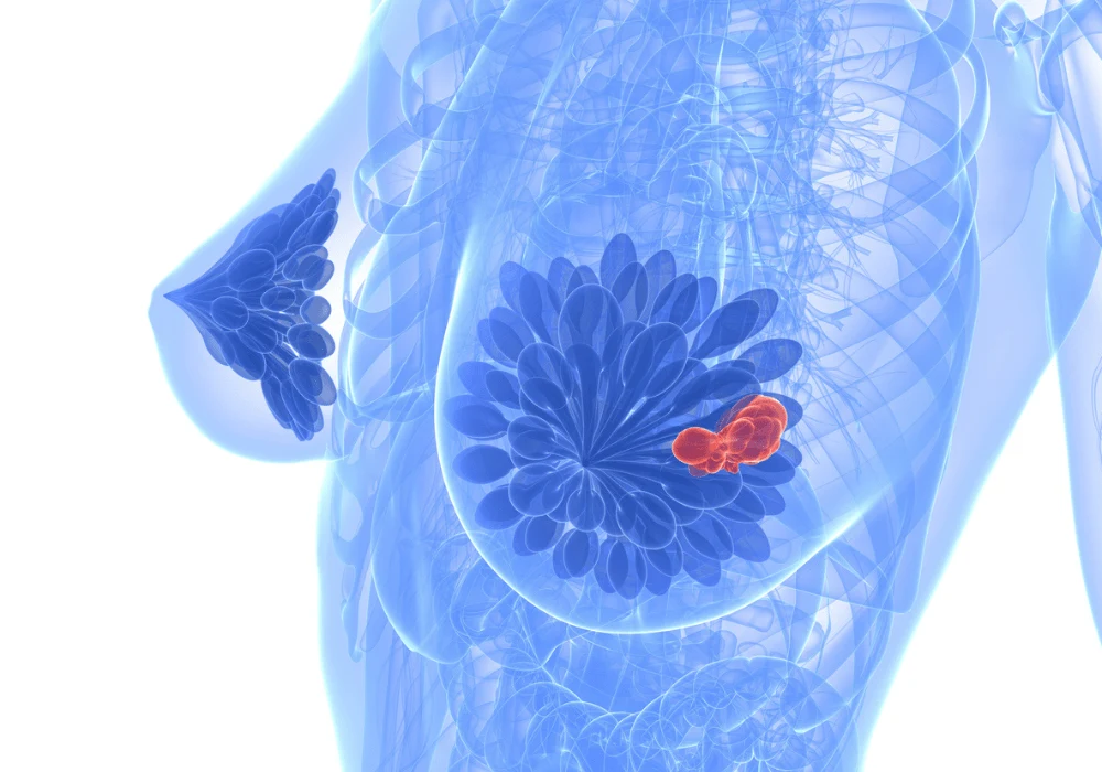
Breast cancer diagnosis and monitoring have long relied on advanced imaging techniques, with magnetic resonance imaging (MRI) playing a critical role. One such technique, diffusion-weighted imaging (DWI), has shown great promise in enhancing breast cancer detection, staging, and monitoring by measuring the diffusion of water molecules in tissues. Recently, further advancements in DWI have emerged, focusing on more sophisticated models like intravoxel incoherent motion (IVIM) and diffusion kurtosis imaging (DKI). However, despite growing research interest, integrating these advanced methods into routine clinical practice remains limited. Recent studies explore the current state of advanced DWI in breast imaging, challenges preventing its wider adoption and the potential future of breast cancer diagnostics.
Role of Advanced Diffusion-Weighted Imaging
Traditional DWI has proven to be an invaluable addition to dynamic contrast-enhanced MRI (DCE-MRI) primarily because it can increase the specificity of cancer detection by evaluating the movement of water molecules within tissues. Using the apparent diffusion coefficient (ADC), the basic model simplifies this motion into a single parameter that reflects free water diffusion and the hindrance caused by tissue microstructures. Although effective, this model only provides a limited view of tissue characteristics.
Advanced DWI techniques go beyond the conventional ADC model. IVIM, for instance, separates molecular diffusion from the pseudo-diffusion related to blood microcirculation. This model allows radiologists to assess both tissue structure and perfusion simultaneously. Meanwhile, DKI captures non-Gaussian diffusion, offering insights into tissue complexity and potentially improving cancer staging. Despite the promise of these techniques, their adoption has been slow, mainly due to the challenges of implementation, which include longer acquisition times and the complexity of data analysis.
Current Application and Obstacles
A recent survey conducted by the European Society of Breast Imaging (EUSOBI) highlights the limited clinical use of advanced DWI in breast imaging. The survey showed that only a small percentage of radiologists actively employ these advanced techniques, with IVIM being the most commonly used but still not widespread. DKI and diffusion tensor imaging (DTI) are even less commonly adopted. This gap between the potential of advanced DWI and its actual clinical use is significant and points to several barriers.
One of the primary challenges is the lack of standardisation in protocols. While standard DWI is relatively straightforward, advanced methods like IVIM and DKI require precise acquisition and data analysis, often involving multiple b-values, which can extend imaging time and increase the complexity of interpretation. Additionally, the availability of user-friendly software for processing and interpreting advanced DWI data is limited, making it difficult for clinicians to implement these techniques efficiently. There is also a need for more robust studies that clearly demonstrate the clinical benefits of advanced DWI over traditional methods in specific diagnostic scenarios.
Advances in Technical Solutions and Research Directions
Several technical advances are being explored to bridge the gap between research and clinical practice. Improved fat suppression techniques and higher-resolution imaging methods have been developed to enhance the accuracy of DWI in breast cancer detection. For example, multi-shot echo-planar imaging (EPI) and simultaneous multislice acquisition techniques have been shown to reduce distortion and improve image quality, making DWI more reliable for small lesion detection.
Another area of focus is reducing acquisition times without sacrificing data quality. Research into optimising b-value selection and minimising the number of required diffusion directions has shown promise. Additionally, deep learning algorithms are beginning to be applied to DWI data analysis, offering potential solutions to the time-intensive process of manually analysing complex datasets. These advancements not only improve image quality but also address the need for faster, more accessible imaging solutions in clinical settings.
The future of breast cancer imaging lies in the successful integration of advanced DWI techniques into routine practice. While current usage remains limited, the potential for these methods to improve cancer diagnosis, staging, and treatment monitoring is immense. Overcoming standardisation, technical complexity, and time efficiency obstacles will be crucial. With ongoing research and collaboration between radiologists, physicists, and industry, the adoption of advanced DWI could revolutionise breast imaging, offering more precise and personalised care for breast cancer patients.
Source: European Radiology
Image Credit: iStock


![Advancing Gastric Cancer Imaging with [18F]ALF-NOTA-FAPI-04 PET/CT Advancing Gastric Cancer Imaging with [18F]ALF-NOTA-FAPI-04 PET/CT](https://res.cloudinary.com/healthmanagement-org/image/upload/f_auto,fl_lossy,q_90,w_200/v1732973927/cw/00128822_cw_image_wi_97a1025129bb4fa62fba8aea90e5883b.webp)

