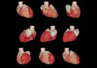
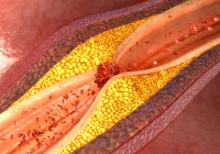
Coronary CT angiography (CCTA) is a widely used non-invasive imaging technique for assessing coronary artery disease. In addition to detecting luminal stenosis, CCTA enables the quantification of atherosclerotic plaque, an important predictor of cardiovascular risk. The accuracy of plaque volume measurement is crucial, as plaque burden, rather than stenosis, is the primary indicator of cardiovascular events. Conventional energy-integrating detector (EID) CT has been the standard for CCTA-based plaque characterisation, but recent advances in imaging technology have introduced photon-counting detector (PCD) CT with ultrahigh spatial resolution (UHR), which offers improved precision in plaque quantification. A recent study published in Radiology evaluates the impact of UHR PCD CT compared with EID CT, specifically examining differences in plaque volume segmentation, reproducibility of measurements and attenuation-based characterisation.
Comparative Analysis of Plaque Quantification
The introduction of UHR PCD CT has led to a significant enhancement in spatial resolution, enabling more detailed assessment of coronary plaque burden. The study found that total plaque volume was consistently lower when measured using UHR PCD CT compared with EID CT. This reduction in total plaque volume was primarily attributed to differences in the segmentation of fibrotic plaque, which appeared significantly lower with UHR imaging. The study also observed that low-attenuation plaque (LAP) volumes were higher in UHR-based measurements. These differences suggest that the improved resolution of PCD CT reduces the effects of partial volume averaging, leading to more refined segmentation of plaque components.
Recommended Read: Ultra-low-dose Coronary CT with Deep Learning Reconstruction
In contrast, calcified plaque volumes did not show significant variation between EID and PCD CT measurements. This finding may indicate that while PCD CT reduces the overestimation of fibrotic plaque, it does not significantly alter the quantification of calcified components. However, a subanalysis of predominantly calcified plaques revealed a slight reduction in calcified volume with PCD CT, suggesting that the enhanced resolution might reduce blooming artefacts commonly associated with calcification in EID CT imaging. These findings emphasise the impact of UHR imaging in providing more precise differentiation of plaque constituents, which could contribute to more accurate cardiovascular risk assessment.
Reproducibility and Reader Agreement
A critical factor in clinical imaging is the consistency and reliability of measurements across different readers. This study demonstrated that UHR PCD CT exhibited excellent reproducibility for total, calcified and fibrotic plaque volumes. Both intra-reader and inter-reader agreement were significantly improved with PCD CT compared to EID CT. The study found that while EID CT showed strong reproducibility for total and fibrotic plaque volumes, its reproducibility for LAP measurements was only moderate to poor. In contrast, PCD CT demonstrated a stronger agreement between repeated measurements and different readers, even for LAP, suggesting that UHR imaging provides a more stable and repeatable method for assessing plaque burden.
This improvement in measurement consistency is particularly relevant for longitudinal studies and clinical decision-making. The ability to reliably track changes in plaque volume over time is essential for evaluating the effectiveness of therapeutic interventions and monitoring disease progression. The study results indicate that the enhanced spatial resolution of UHR PCD CT minimises variability in plaque segmentation, making it a more dependable tool for clinical and research applications.
Plaque Attenuation and Histogram-Based Characterisation
Beyond volume-based assessments, the study also analysed the attenuation characteristics of plaques using histogram-based analysis. It was observed that PCD CT produced significantly higher median attenuation values than EID CT, alongside a wider dynamic range of attenuation measurements. Additionally, the variance in attenuation values was greater in PCD CT imaging, indicating that UHR provides a more detailed and nuanced differentiation between different plaque components.
These differences in attenuation values suggest that the improved resolution of PCD CT may enhance the accuracy of tissue characterisation within plaques. The broader dynamic range of attenuation values could allow for a more precise classification of fibrotic, low-attenuation and calcified plaque components. However, the study highlights the need for further validation of attenuation thresholds specific to UHR imaging. Current thresholds used for plaque characterisation were originally established using EID CT, and their direct application to PCD CT may not fully capture the improved resolution and sensitivity of UHR imaging. Future research should focus on establishing UHR-specific attenuation thresholds to optimise plaque classification and ensure consistency across different imaging technologies.
The findings of this study highlight the advantages of UHR PCD CT in coronary plaque quantification. The reduction in total plaque volume observed with PCD CT suggests that the improved spatial resolution reduces the overestimation of fibrotic plaque while providing more accurate segmentation of LAP. Additionally, the study demonstrated that PCD CT offers significantly improved reproducibility, particularly for LAP measurements, which have historically been less reliable using EID CT. The improved inter-reader agreement suggests that UHR PCD CT provides a more stable and repeatable method for assessing plaque burden, making it a valuable tool for both clinical practice and research.
The increased attenuation values and wider dynamic range observed with PCD CT underscore the potential of UHR imaging for improved plaque characterisation. However, further research is needed to define attenuation thresholds specific to PCD CT, ensuring optimal classification of plaque components. These advancements in imaging technology have the potential to refine cardiovascular risk stratification and improve the accuracy of non-invasive coronary plaque assessment, ultimately contributing to better patient management and treatment strategies.
Source: Radiology
Image Credit: iStock

