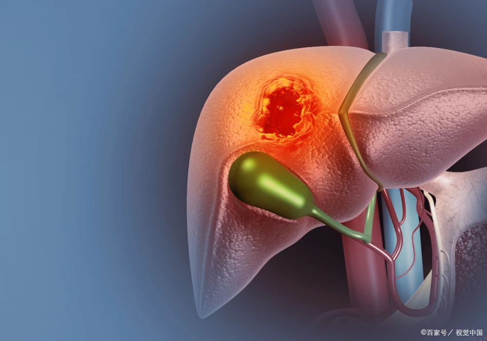
Hepatocellular carcinoma (HCC) presents with marked biological diversity that shapes prognosis, treatment sensitivity and risk of recurrence. Molecular and pathological classifications capture this heterogeneity but usually rely on tissue sampling that is invasive, prone to sampling error and often unavailable when therapy is chosen. Increasing evidence shows that multiphase computed tomography (CT) and multiparametric magnetic resonance imaging (MRI) reveal patterns that align with recognised molecular classes and histological subtypes. Interpreting these features can support non-invasive phenotyping, guide biopsy decisions and inform therapeutic thinking. Although correlations are imperfect and validation remains incomplete, an integrated approach that combines imaging with clinical, pathological and molecular data is emerging as a practical route to more individualised HCC management.
Linking Imaging with Biological Classes
At a molecular level, HCC is broadly divided into proliferative and non-proliferative classes. The proliferative class, which represents about half of cases, is associated with aggressive behaviour, higher alpha-fetoprotein and des-gamma-carboxy prothrombin levels. Non-proliferative tumours tend to retain hepatocyte-like features, show higher chromosomal stability and more frequent promoter mutations and are more often linked to alcohol-related steatotic liver disease, metabolic dysfunction-associated steatohepatitis and hepatitis C virus infection. Imaging can reflect aspects of these classes: proliferative tumours more often display features linked to invasion and necrosis, whereas non-proliferative patterns include intratumoral fat and uptake characteristics that track with β-catenin activation. Because one-to-one mapping is rare and overlap exists within and between lesions, current best practice treats imaging as a proxy that complements, rather than replaces, tissue and molecular assessment.
Patterns That Suggest Specific Subtypes
Two pathological subtypes commonly align with the proliferative class: macrotrabecular-massive HCC (MTM-HCC) and progenitor-phenotype HCC. MTM-HCC, seen in a minority of cases, is linked to larger size, higher grade, microvascular invasion, satellite nodules and poorer outcomes. On CT/MRI it is frequently associated with intratumoral necrosis or hypovascular components on the arterial phase, rim arterial phase hyperenhancement, intratumoral arteries, peritumoral (corona) enhancement, non-smooth margins and, at times, tumour in vein. Lower apparent diffusion coefficient values and larger dimensions further support suspicion. Several scores and radiomics models that incorporate necrosis, haemorrhage and diffusion metrics have shown promising discrimination, though none is sufficiently validated for routine clinical application.
Internal link: MRI Habitat Patterns Flag MVI Risk and Prognosis in HCC
Progenitor-phenotype HCC, defined by expression of markers such as cytokeratin 19 or EpCAM, shows an infiltrative growth pattern, frequent vascular invasion and worse disease-free survival after resection. On gadoxetic acid-enhanced MRI and diffusion-weighted imaging, recurrent cues include non-smooth margins, rim hyperenhancement in the arterial phase, targetoid appearances recognised by LI-RADS, lower tumour-to-liver ratios on diffusion and hepatobiliary phase images and absence of mosaic architecture. Quantitative texture features and radiomics have improved prediction in research cohorts but, as with MTM-HCC tools, require external validation across diverse populations and imaging protocols.
Among non-proliferative tumours, steatohepatitic HCC (SH-HCC) typically shows intratumoral fat and is often smaller than other subtypes. In metabolic dysfunction-associated backgrounds, intratumoral fat without mosaic architecture can be highly specific, although fat is not exclusive to this subtype and may be difficult to assess against a steatotic liver. Quantitative fat fraction techniques can help but may miss small foci. Notably, the distribution of fat within the lesion may carry prognostic information, with homogeneous fat patterns linked to smaller size and, in some series, favourable outcomes.
CTNNB1-mutated HCC constitutes a frequent molecular subtype, with canonical WNT/β-catenin activation and an immune-cold microenvironment that has been associated with reduced benefit from immune checkpoint inhibitors. On gadoxetic acid-enhanced MRI, these tumours can appear iso- or hyperintense on the hepatobiliary phase and may show higher diffusion metrics, reflecting preserved or enhanced uptake transporter expression. Radiomics signatures derived from hepatobiliary and portal venous phase images have reported encouraging accuracy for mutation status estimation, yet sample sizes have been modest and external validation limited.
Current Limitations and Priorities for Validation
Despite consistent associations, imaging features seldom map uniquely to a single molecular or pathological category. Many studies rely on histology as a proxy for genomics, even though these domains only partially overlap. Within-tumour and between-tumour heterogeneity further complicate correlation, and current classifications rarely accommodate lesions that exhibit mixed proliferative and non-proliferative attributes. Most evidence to date comes from single-centre retrospective cohorts with small samples and a predominance of chronic hepatitis B virus infection, which constrains generalisability to broader aetiologies, including metabolic disease. Methodological challenges also persist, such as inter-reader variability in estimating necrotic or hypovascular components and the difficulty of precise radiology–pathology spatial matching. Although artificial intelligence-assisted mapping and patient-specific 3D moulds offer a potential solution, they remain research tools rather than everyday practice.
CT and MRI provide practical, non-invasive markers that align with recognised molecular classes and histological subtypes of HCC, supporting risk stratification, treatment planning and biopsy triage. Proliferative-pattern imaging has been linked with lower transarterial chemoembolisation response and may point towards combined tyrosine kinase inhibitor and immune checkpoint inhibitor strategies, whereas hepatobiliary phase characteristics and intratumoral fat can suggest non-proliferative biology. Because no single model is ready for routine use and correlations are imperfect, the most robust path forward is an integrated phenotyping framework that unites clinical data with imaging, pathology and molecular profiling. Larger, multi-centre prospective studies with rigorous radiology–pathology–genomics correlation are needed to confirm utility and embed imaging-informed subtyping into everyday decision-making.
Source: European Radiology
Image Credit: iStock


