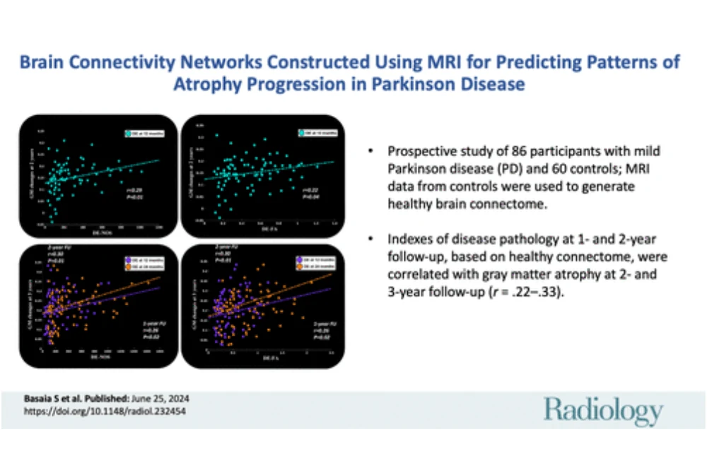
Parkinson’s disease (PD) is a neurodegenerative disorder marked by motor symptoms like bradykinesia, rigidity, resting tremor, and gait disorders, alongside nonmotor symptoms such as cognitive impairment, REM sleep behaviour disorder, and hyposmia. The accumulation of misfolded α-synuclein protein within Lewy bodies and neurites signifies PD progression, spreading trans-synaptically through brain regions. Understanding how brain architecture influences this spread remains challenging. , recently published in Radiology, explores how graph analysis and MRI connectomics provide insights into structural and functional connectivity (SC and FC) in the brain, potentially predicting the progression of grey matter (GM) atrophy in mild PD patients.
Graph Analysis and Brain Connectivity in PD
Graph-based network analysis portrays the brain as a network of nodes (brain regions) and edges (connections), offering a comprehensive view of disease-induced changes in brain organisation. This method has revealed that the SC and FC between brain regions significantly influence the spatial spread of PD pathology. For instance, regions with strong SC and FC to already affected areas may exhibit greater atrophy over time. By mapping these connections, researchers can predict clinical progression from the early disease stages, aiding in understanding the neurodegeneration process and hypothesising about disease mechanisms.
Study Design and Methodology
The current study aimed to assess the SC and FC of brain regions in healthy controls and relate this to GM atrophy in patients with mild PD. Eighty-six mild PD patients and 60 age- and sex-matched healthy controls were recruited. Over a three-year follow-up, participants underwent clinical, cognitive, and MRI assessments. Disease exposure (DE) indexes, calculated based on the SC and FC in healthy controls and the severity of atrophy in PD patients, were used to predict atrophy progression. The study focused on how baseline connectivity in healthy brains might relate to subsequent atrophy in PD-affected brains, providing a predictive model for disease progression.
Results and Findings
The study found significant correlations between DE indexes at one- and two-year follow-ups and GM atrophy at subsequent time points. Regions with higher SC and FC with atrophic areas in PD patients showed more significant atrophy over time. Specifically, the right caudate nucleus, and temporal, parietal, and frontal regions were early targets of atrophy. These findings align with previous studies indicating these areas are vulnerable in early PD stages. The study also observed left hemispheric lateralisation of cortical thinning, consistent with existing literature on hemisphere susceptibility in neurodegenerative diseases.
Discussion and Implications
Graph analysis and MRI connectomics have shown that structural and functional brain architecture can predict GM atrophy progression in PD. The observed relationship between SC and FC suggests that structural and functional effects are interrelated in the disease spread. This predictive capability is crucial for early diagnosis, understanding disease mechanisms, and developing targeted treatments. Future research should explore patient-specific connectivity changes and integrate other factors like gene expression and molecular imaging to deepen the understanding of PD progression and its underlying mechanisms.
This study demonstrates that the brain's structural and functional connectivity can play a critical role in predicting the progression of GM atrophy in mild Parkinson’s disease. Utilising disease exposure indexes based on healthy brain connectomes offers a promising approach for early identification of atrophy, enhancing prognostic accuracy, and informing therapeutic strategies. Further research with extended follow-ups and additional imaging techniques will be vital to refine these predictive models and improve PD management. Understanding the interplay between brain connectivity and neurodegeneration could lead to breakthroughs in combating this debilitating disease.
Source & Image Credit: Radiology


