Radiology is at the forefront of a digital revolution, with artificial intelligence (AI) transforming traditional diagnostic workflows. Among the most significant advancements is the application of large language models (LLMs) like OpenAI’s GPT-4. Equipped with its Advanced Data Analysis (ADA) capabilities, GPT-4 has the potential to aid radiologists in overcoming longstanding challenges. Radiology faces a shortage of professionals with strong statistical knowledge, which can limit the adoption and critical engagement with AI tools. A recent article published in Radiology explores the capabilities of GPT-4 ADA in the evaluation of chest radiographs, demonstrating how this technology can support radiologists in tasks ranging from data visualisation to machine learning model development.
Barriers to AI Integration in Radiology
While AI’s benefits in radiology are becoming increasingly evident, integrating these tools within clinical settings has not been without difficulties. Financial limitations, IT integration challenges and validation issues remain persistent obstacles to widespread AI adoption. Additionally, many radiologists lack formal training in statistical methods, essential for understanding and verifying AI-generated insights. This gap not only impedes the integration of AI but also restricts the critical assessment of AI tools, often leaving radiologists to rely on AI outputs without a full understanding of their statistical underpinnings.
Moreover, radiologists are faced with the balancing act of incorporating clinical evidence with the commercial interests of AI providers, further complicating the path to effective AI adoption. Here, GPT-4 ADA presents a unique opportunity. As a powerful LLM capable of natural language processing, reasoning and data analysis, GPT-4 ADA offers a simplified yet effective interface that could empower radiologists to autonomously conduct data analyses without needing extensive statistical or programming knowledge. By democratising AI analysis, GPT-4 ADA promises to bridge the technical gap that has long hindered AI adoption in radiology.
GPT-4 ADA’s Application in Chest Radiograph Evaluation
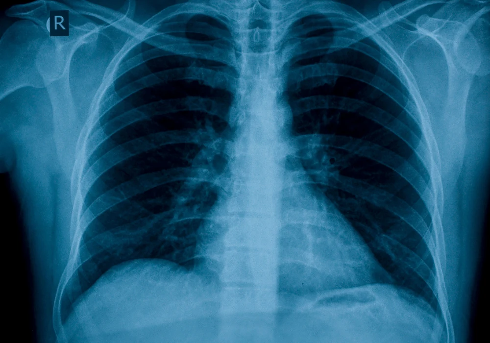
In a recent study, researchers assessed GPT-4 ADA’s performance on a dataset of bedside chest radiographs from University Hospital RWTH Aachen. The model was tasked with various analytical functions, including plotting radiograph usage trends, performing descriptive statistical analyses and setting up predictive models to identify pulmonary opacities. The study involved 43,788 chest radiograph reports, accompanied by demographic and laboratory data from patients in intensive care, creating a complex dataset that would typically demand advanced technical expertise to analyse effectively.
GPT-4 ADA autonomously executed these tasks, generating insights comparable to those developed by specialist models. The AI model was able to visualise radiograph usage trends over time and determine statistical associations within the dataset, providing both descriptive and quantitative insights. For predictive modelling, GPT-4 ADA implemented gradient boosting techniques and logistic regression models to forecast pulmonary opacity occurrences, achieving an area under the curve (AUC) of 0.75, closely matching the 0.80 AUC achieved by human specialists.
Despite minor statistical inaccuracies, GPT-4 ADA demonstrated consistency and reliability in its outputs, offering radiologists an accessible tool for robust data analysis. Researchers validated the AI-driven analyses, which were largely accurate, with performance metrics, such as sensitivity and specificity, comparable to those produced by experts. Notably, GPT-4 ADA’s output accuracy was maintained even when operating without explicit guidance, highlighting its potential for autonomous analysis in real-world settings where clinician involvement might be limited.
Transforming Radiology Through Accessible AI Solutions
The successful application of GPT-4 ADA in chest radiograph analysis has far-reaching implications for the future of AI in radiology. In the future, LLMs could become integral tools in routine diagnostic processes, enabling radiologists to shift their focus from technical data manipulation to clinical decision-making. GPT-4 ADA can reduce the dependency on specialised data science skills by providing a user-friendly interface capable of autonomous analysis, encouraging wider AI adoption across radiology departments.
However, the journey towards AI-driven radiology must be balanced with rigorous validation to ensure accuracy and reliability in clinical contexts. The deployment of LLMs like GPT-4 ADA will require ongoing evaluation to mitigate potential errors and inconsistencies. As demonstrated in the study, GPT-4 ADA’s outputs were not flawless, with minor statistical inaccuracies underscoring the need for continuous monitoring and refinement. Nevertheless, its performance signals a transformative shift in radiology, where AI-driven tools could soon play a pivotal role in enhancing diagnostic accuracy, efficiency and accessibility.
GPT-4 ADA’s performance in this study underscores its potential as a valuable tool for radiologists, offering a pathway towards more efficient, data-driven chest radiograph evaluation. With its intuitive interface and advanced analytical capabilities, GPT-4 ADA bridges the knowledge gap in statistical and data science skills, empowering radiologists to make informed decisions based on reliable AI-driven insights. The future of diagnostic analysis may well lie in accessible LLMs that blend technological sophistication with user-centric design, ultimately enhancing patient outcomes through data-informed diagnostics.
Source: Radiology
Image Credit: iStock

