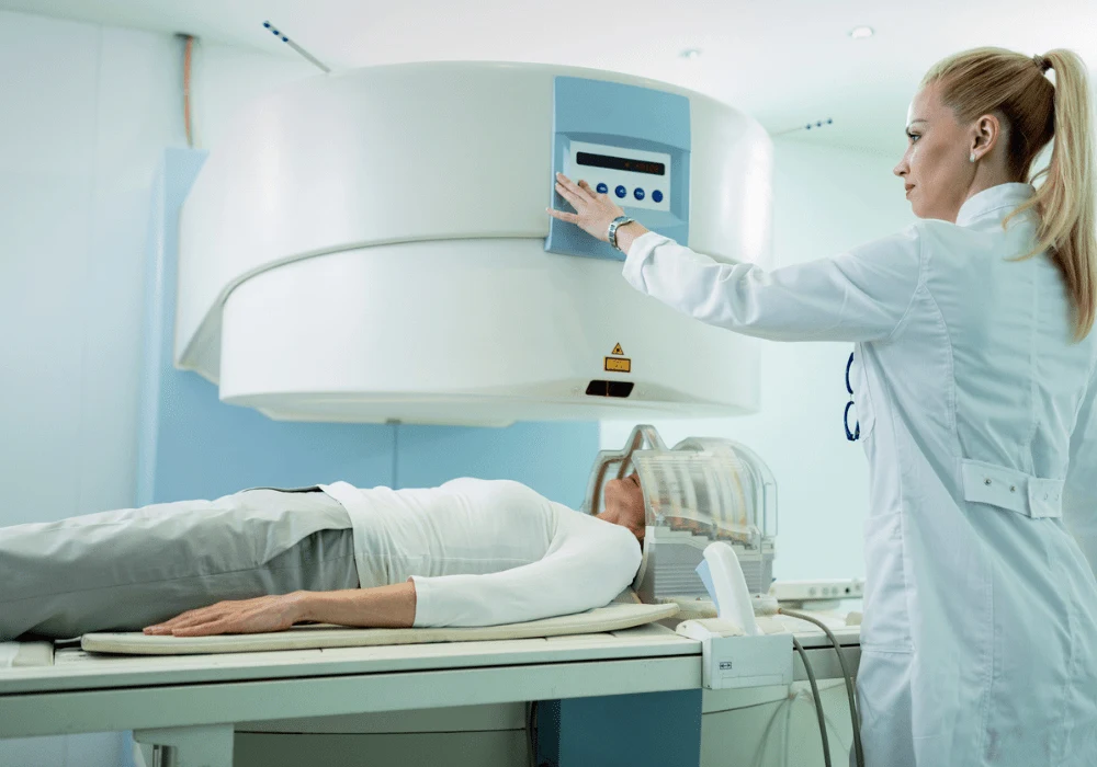
Accurate distinction between benign and malignant breast lesions is critical for guiding patient management and avoiding unnecessary interventions. Traditional imaging methods, while effective for initial detection, often lack sufficient specificity, resulting in reliance on biopsies. Recent advances in MRI techniques offer new opportunities to improve diagnostic precision. Time-dependent diffusion MRI (Td-dMRI), particularly using the IMPULSED approach, enables quantification of tissue microstructure, which may enhance lesion characterisation. A recent study published in Radiology: Imaging Cancer explores the diagnostic value of microstructural metrics derived from IMPULSED Td-dMRI in differentiating breast lesion types, comparing their performance with conventional apparent diffusion coefficient (ADC) metrics from diffusion-weighted imaging (DWI).
Microstructural Differences in Benign and Malignant Lesions
Histologically, malignant breast tumours tend to exhibit increased cell size, higher cell density and greater intracellular volume fraction compared to benign counterparts. Td-dMRI allows noninvasive assessment of these features by estimating cell diameter, cell density, intracellular volume fraction (Vin) and extracellular diffusivity (Dex). The IMPULSED technique integrates oscillating and pulsed gradient spin-echo sequences to sample diffusion over varying timescales, enabling detailed mapping of tumour microarchitecture.
Must Read: Differentiating Benign and Malignant Breast Lesions with Ultrafast MRI
In this prospective study, participants underwent both conventional DWI and Td-dMRI prior to biopsy or surgery. Quantitative metrics were extracted from Td-dMRI maps and compared with pathologic outcomes. Malignant lesions demonstrated significantly higher cell diameter, cell density and Vin than benign lesions, whereas ADC values were significantly lower in malignancies. Dex did not differ notably between groups. These findings reflect the proliferative and structural characteristics typical of malignant tissue and suggest that Td-dMRI metrics provide a more direct assessment of underlying cellular properties than ADC.
Diagnostic Accuracy of IMPULSED Td-dMRI Metrics
The diagnostic performance of each diffusion metric was assessed using receiver operating characteristic analysis. Among all metrics, cell density derived from Td-dMRI achieved the highest area under the curve (AUC) at 0.93, outperforming ADC, which recorded an AUC of 0.79. Cell diameter and Vin showed moderate diagnostic value, with AUCs of 0.73 and 0.82, respectively. Dex had limited diagnostic utility, with an AUC of 0.58.
Cell density also achieved the highest positive predictive value of 98% at an optimal threshold, making it particularly useful in reducing false-positive diagnoses. Notably, sensitivity and specificity were also high, indicating robust performance across lesion types. By directly measuring the number of cells in a given volume, cell density offers a reliable marker of malignancy. This advantage positions Td-dMRI as a superior technique to conventional DWI in differentiating lesion types, with potential implications for reducing reliance on invasive diagnostic procedures.
Clinical Implications and Future Directions
The integration of IMPULSED Td-dMRI into clinical breast imaging protocols could significantly improve diagnostic accuracy, particularly in cases where conventional imaging yields inconclusive results. The ability to noninvasively characterise tumour microstructure aligns with a broader shift toward precision medicine. While the acquisition time for Td-dMRI remains relatively long and image analysis complex, advancements in imaging technology and artificial intelligence may address these limitations, making the approach more feasible for routine clinical use.
Beyond breast cancer, Td-dMRI metrics have demonstrated diagnostic utility in other malignancies, including prostate, ovarian and paediatric brain tumours. Their application in breast imaging thus represents a logical extension of a proven methodology. Future studies involving larger, multi-centre cohorts could further validate the reproducibility and generalisability of these findings. Moreover, exploration of Td-dMRI metrics in the context of tumour grading and molecular subtyping may expand their role in personalised oncology.
The study underscores the potential of IMPULSED Td-dMRI as a noninvasive imaging tool to differentiate benign from malignant breast lesions. Among the evaluated metrics, cell density emerged as the most effective biomarker, outperforming traditional ADC measures. By capturing the fundamental microstructural differences between lesion types, Td-dMRI may reduce the need for unnecessary biopsies and support more accurate, early-stage cancer diagnosis. With further validation and technical refinement, microstructural imaging could become a cornerstone of advanced breast cancer diagnostics.
Source: Radiology: Imaging Cancer
Image Credit: iStock


