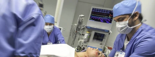
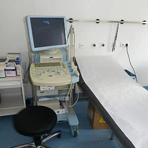
With growing interest in understanding muscle atrophy and function in critically ill patients and survivors, ultrasound is emerging as a potentially powerful tool for skeletal muscle quantification. However, there are key challenges that need to be addressed in future work to ensure useful interpretation and comparability of results across diverse observational and interventional studies, according to a research paper published in Annals of the American Thoracic Society.
"Ultrasound presents several methodological challenges, and ultimately muscle quantification combined with metabolic, nutritional, and functional markers will allow optimal patient assessment and prognosis," the report authors say. "Moving forward, we recommend that publications include greater detail on landmarking, repeated measures, identification of muscle that was not assessable, and reproducible protocols to more effectively compare results across different studies."
Features of skeletal muscle (including muscle quantity measures like mass and cross-sectional area and muscle quality measures such as architecture and evidence of myonecrosis) may provide a more feasible and objective approach to assessing muscle health in ICU patients. Objective quantifications of muscle (which include, but are not limited to, muscle mass, thickness, and cross-sectional area) that are sufficiently sensitive to detect small changes over acute timeframes may ultimately facilitate evaluation of interventions to counter muscle atrophy and weakness.
Over the last decade, common imaging technologies, such as ultrasound and computed tomography (CT) have been translated into the ICU for quantification of muscle. The transformational application of these techniques, the report says, has advanced our understanding of the muscle characteristics of ICU patients at admission and during the ICU trajectory. Quantification of skeletal muscle, an example of this transformational application, allows for identification of those with low muscle quantities. Low muscle quantities, in turn, have been related to poor clinical outcomes including increased mortality, as well as reduced ventilator-free days and ICU-free days.
Notably, the technique of critical care skeletal muscle ultrasound lags behind other critical illness research domains where standardisation of definitions, gradation of severity and technical skills have allowed assessment of external validity of data produced.
While ultrasonography is an attractive tool for evaluating muscle health, critical care muscle ultrasound research is hampered by a number of factors including:
- No formal training programme or standardised protocol used to educate clinicians, healthcare providers or students
- Lack of standardisation in terms of specific muscles and muscle groups to be analysed
- The number of images needed for adequate analysis of biomechanical measures and metabolic integrity also remains unstandardised
- Variability in the data that is collected across studies (i.e., muscle layer thickness, cross-sectional area, echogenicity or a combination of these and other measurements)
- There are a limited number of studies that provide a healthy, homogeneous cohort for comparison
"While ultrasound presents a potential option to measure muscle mass in real-time in clinical facilities, we need to first identify and validate a universal protocol that can be easily implemented in the critical care setting to enhance and advance studies that aim to improve muscle rehabilitation in survivors of critical illness," the authors conclude.
Source: Annals of the American Thoracic Society
Image Credit: Pixabay
References:
Mourtzakis, Marina et al. (2017) Skeletal Muscle Ultrasound in Critical Care: A Tool in Need of Translation. ATS Journals. doi.org/10.1513/AnnalsATS.201612-967PS
Latest Articles
muscle atrophy, skeletal muscle ultrasound, muscle quantification
With growing interest in understanding muscle atrophy and function in critically ill patients and survivors, ultrasound is emerging as a potentially powerful tool for skeletal muscle quantification. However, there are key challenges that need to be addres


