


ICU Management & Practice, ICU Volume 12 - Issue 3 - Autumn 2012
In this article we look at strategies for preventing lower airway colonisation, focusing on the factor that is widely-believed to be the main culprit forventilator associated pneumonia (VAP) development: the endotracheal tube (ETT).
VAP and its Pathogenesis
Ventilator associated pneumonia, defined as occurrence of pneumonia at least 48–72 hours after commencement of mechanical ventilation, is known to be associated with increased hospital stay and costs (Chastre 2002). However, its impact on mortality is currently under debate (Bekaert et al. 2011; Forel et al. 2012).
The main cause for VAP
development is currently believed to be the ETT, rather than the ventilator.
The presence of an ETT is an independent risk factor for developing VAP. The
ETT disrupts the cough reflex, promotes accumulation of tracheobronchial secretions
and microbial biofilm, and provides a direct conduit for pathogenic
microorganisms to reach the lower respiratory tract by gravity draining through
micro-channels around the cuff, and by gravity detaching from the inner lumen.
The ETT acts thereby as a bridge for oropharingeal pathogens to colonise the
lower airways, potentially leading to an infectious process. Several strategies
to prevent VAP have been developed, targeting the role of the ETT as the main
pathogenic factor involved in its development.
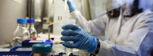
Preventing Biofilm Build-Up
Soon after intubation, a thin layer of biofilm is present on the
inner surface of the ETT. During mechanical ventilation, biofilm thickness
increases because secretions tend to accumulate inside the ETT, thus reducing the
volume available for airflow, despite the regular use of a suctioning catheter.
Aggregates of biofilm pathogens can be detached via suctioning maneuvers or
simply via mechanical ventilation; these then reach the lower airways. Data
suggest that the same pathogens present in the ETT biofilm cause lower
respiratory tract colonisation, leading to VAP (Gil-Perotin et al. 2012).
Different strategies have been employed to reduce or remove biofilm inside the
ETT by means of anti-bacterial drug coatings or cleaning devices.
Coating
The ETT can be coated with a large variety of substances; however, only two coatings have been tested in clinical trials. In a trial involving 46 ICU patients who were randomised to be intubated with either a standard or coated ETT, silversulfadiazine coating proved to be effective in preventing ETT bacterial colonisation (Berra et al. 2008). Furthermore, in a large randomised trial by Kollef and colleagues, involving more than 1,500 patients, the use of silver-coated ETTs reduced the incidence of microbiologically proven VAP (Kollef et al. 2008). Silver is effective because its ions penetrate inside the pathogen’s membrane and interfere with nucleic acids replication, preventing proliferation.
An alternative approach to inducing biofilm pathogens’
cell death is topical photosensitisation. This technique is based on a
photosensitising agent (methylen blue plus photoreacting agent) and a light diffuser
catheter that activates the agent, leading to formation of oxygen radicals. Though
photosensitisation has been studied only in vitro, it has shown promising results
in reducing bacterial survival of P. Aeruginosa and Methicillin resistant S. Aureus
strains (Biel et al. 2011; Berral et al. 2008).
Another approach involves changes in material topography in an
attempt to limit pathogen adhesion. Pathogens easily adhere and form biofilms
on the hydrophobic polyvinylchloride (PVC) surface, a standard ETT. The
chemical treatment of a PVC surface forms roughness at the nanometer level,
resulting in reduced surface hydrophobicity and reduced bacterial adhesion (Loo
et al. 2012). In vitro results have shown the combination of nanorough ETT and
the presence of a fructose coating is effective in decreasing biofilm formation
(Durmus et al. 2012). Although promising, the efficacy and safety of these
techniques still need to be tested in the clinical setting.
Cleaning the ETT
The physical removal of biofilm from the inner lumen of the ETT was proposed by Kolobow and colleagues in 2005 (Kolobow et al. 2005). A dedicated device called the Mucus Shaver, consisting of an inflatable balloon with rubber rings embedded, proved to be effective in removing biofilm from ETTs used in animal models. Recently, a randomised trial showed the safety and feasibility of the use of the Mucus Shaver in a clinical setting bacterial growth and biofilm thickness were reduced in the ETTs of treated patients (Berra et al. 2012). While the Mucus Shaver is not commercially available, other devices with intended similar use are present on the market (Rescue Cath from Omneotech, a cleaning device from endOclear LLC, and a closed suction system from BIOVO Technologies). Whether or not physical biofilm removal is effective in VAP prevention still needs to be assessed; however, the importance of biofilm removal might also extend to other means, such as its relevance in maintaining the antibacterial properties of a coated ETT (Berra et al. 2006). Clinical studies are needed to assess whether coating and regular biofilm removal act synergistically.
Preventing Drainage From the Oropharynx
Other than via the presence of biofilm, a method in which pathogens can reach distal airways is through drainage from the hypopharynx. Standard ETTs are equipped with high-volume, low-pressure PVC cuffs, which fold against the tracheal wall forming micro-channels between the hypopharynx and the subglottic space. Maintenance of cuff internal pressure is crucial to preventing microaspirations, and continuous control through a dedicated device is effective in reducing the drainage of gastric contents into the airways (Nseir et al. 2011). Although the pressure of the ETT cuff is adequate, secretions drain towards the subglottic space. Several strategies have been proposed to prevent secretion drainage, such as subglottic secretion drainage (SSD) systems, cuff modifications to limit the presence of micro-channels and removal and anti-gravitational positioning to prevent the movement of secretions from the ETT and the hypopharynx into the lower airways.
Subglottic Drainage
SSD systems usually consist of a small accessory lumen on the ETT, with a subglottic opening, connected to a negative-pressure generator. The material present above the ETT cuff in the subglottic space is removed, continuously or intermittently, through the accessory lumen. A randomised clinical trial on more than 300 ICU patients showed a reduction in VAP incidence in the group treated with a SSD system (Lacherade et al. 2010); furthermore, a recent meta-analysis confirmed the efficacy of SSD systems in VAP prevention (Muscedere et al. 2011).
A novel approach for subglottic drainage has been developed by BIOVO
Technologies. The Airway Medix endotracheal tube is equipped with a
self-expanding sleeve sealed to the tracheal mucosa, while a collecting basin
built into the sleeve provides safe drainage of pooled subglottic secretions.
Another strategy to removing secretions is the Mucus Slurper (Kolobow et al.
2006), which consists of a modified ETT, equipped with a series of small
suctioning channels opening at the tip. An aspiration system removes through
the suctioning channels both secretions leaked through the cuff and the biofilm
present on the inner lumen of the ETT. No clinical data are currently available
about the efficacy of the Mucus Slurper.
Cuff Material and Shape Modifications
Many materials have been proposed to substitute PVC cuffs, such as poliurethane (PU), lycra and latex, which have demonstrated better sealing in vitro. The use of materials other than PVC should reduce the formation of microchannels, resulting in reduced secretion drainage. However, in the clinical setting, only PU cuffs have been evaluated. The use of PU-cuff equipped ETTs resulted in lower postoperative pneumonia in a population of cardiac surgery patients (Poelaert et al. 2008). The combination of PU cuff and SSD was effective in reducing VAP in a clinical randomised trial (Lorente et al. 2007); however, it is not clear how much of the preventive effect is attributable to the use of a PU cuff. Another approach to limiting the folding of the cuff surface is to modify the shape of the cuff using a tapered shape rather than the classical cylindrical one. Although ETTs equipped with tapered cuffs resulted in better sealing in vitro, the clinical effect of this approach on VAP prevention still needs further investigation.
Positioning
A novel approach that has been proposed to reduce the movement of pathogens towards the distal airways is the adoption of the lateral head-down position, otherwise known as the lateral Trendelenburg position. This involves maintaining the main axis of the trachea slightly below the horizontal plane, so that gravity will favour drainage of secretions outside the airways. In animal models, the head-down position resulted in absence of bacterial colonisation of the airways, while those in the control group were heavily colonised (Panigada et al. 2003). Moreover, mucus flow inside the ETT was dependent on gravity, being directed towards the lungs in the standard head-up position, and towards the ventilator circuit in the head-down position (Li Bassi et al. 2008). These preclinical studies are the basis for a currently ongoing clinical trial, the Gravity-VAP Trial, which compares the lateral head-down position with the standard semirecumbent position, with the aim of disclosing a new preventive measure for VAP. This project is endorsed by the European Critical Care Research Network of the European Society of Intensive Care Medicine (ESICM).
Conclusions
A passage of pathogens through the outside or the inside of the ETT, which are under gravitational forces, has been proposed as a major offender of the lungs during mechanical ventilation. Several intriguing preventive strategies have focused on interrupting the bridge of secretions or biofilm from the oropharyngeal cavity to the lower respiratory tract. Larger clinical studies need to be performed to evaluate the benefits of such novel strategies for our patients.