


ICU Management & Practice, ICU Volume 12 - Issue 2 - Summer 2012
Ultrasonography with a head-to-toe approach has become a comprehensive tool in evaluating critically ill patients from the bedside. In this article, authors propose an innovative approach to critical care ultrasonography, which is simple, fast and transferrable to different anatomical sites.
Introduction
In the past decade many authors have emphasised the role of ultrasonography in daily patient assessment (Beaulieu and Marik, 2005a;b), even proposing to replace, rather than accompany, the traditional stethoscope. A particular role for ultrasonography is emerging in the management of emergency room (Gunst et al. 2008) and ICU patients (Kendall et al. 2004), where its advantages are well known, including, but not limited to, the absence of radiation exposure, and cost-effectiveness (Kendall et al. 2007). For the critical care patient, instead of concentrating on a specific anatomical district, ultrasonography seems to evolve in the direction of a totalbody approach (Karabinis et al. 2010).
Critical Care Ultrasonography (CCU) is emerging as a multidisciplinary technique, where morpho-functional information, obtained with imaging in different anatomical districts, is integrated with clinical data in real-time by the intensivist (Bouhemad et al. 2007). Beyond already mentioned advantages, CCU permits 24-hour bedside availability and repeatability (Harding et al. 2011), rapid information accumulation on all anatomical districts, possibility to provide assistance to invasive manoeuvres (Hind et al. 2003; Sustic et al. 2000) and titration of mechanical ventilation (Luecke et al. 2012).
Why is CCU Different?
In the ICU or emergency department, where CCU is performed, the irregularity of the setting clearly discloses major differences in both patient approach and physician observation compared with those in traditional ultrasonography, performed by the radiologist.
The physical approach to the patient is somewhat unstructured: the physician may prefer not to follow an anatomical order, giving priority to ruling out potentially lifethreatening conditions (Nagdev and Stone, 2011), assessing the function of vital organs (Jambrik et al. 2004), or looking for early signs of organ failure (Corradi et al. 2011). This subversion from the classical head-totoe physical exam recalls that of Advanced Life Support, where priority is given to the identification of clinical situations that might precipitate a patient’s conditions.
CCU is held at a clinical moment where a functional evaluation, rather than anatomical, is needed, since it consists of a problemfocused analysis that gives a multidisciplinary visual. Given the emerging relevance of CCU, the intensivist must focus his/her attention on using such tool appropriately; simple questions must be posed and simple answers sought. The main intent of CCU is to allow early goal-directed therapy.
The approach we are trying to propose is conceptual rather than practical, and the aim is to give the physician a short list of questions to pose each time the probe touches a specific part of the body. The simplicity of the approach we focus on does not want to be reductive and we share the invite of other authors (Vignon et al. 2007) for others to be involved in promoting continuous formation in ultrasonography for intensivists.
The WAMS-D Approach
Authors of this paper want to propose a generic approach that can adapt to different situations, involving disparate anatomical sites. Without pretending to give a universal approach, we suggest a conceptual flow that can help in disentangling complex questions that crowd the physician’s mind once a probe is placed on the patient’s skin.
To promote this fluidity, questions and answers have only one mandatory characteristic: simplicity. Yes/no questions are the most valuable, especially if answers are given in a reasonable timespan. The algorithm that follows focuses on the observation that ultrasonography can rapidly identify water and liquids (black), air (barrier effect), see moving parts and measure distances. This is all related to water, air, movement, size and Doppler.
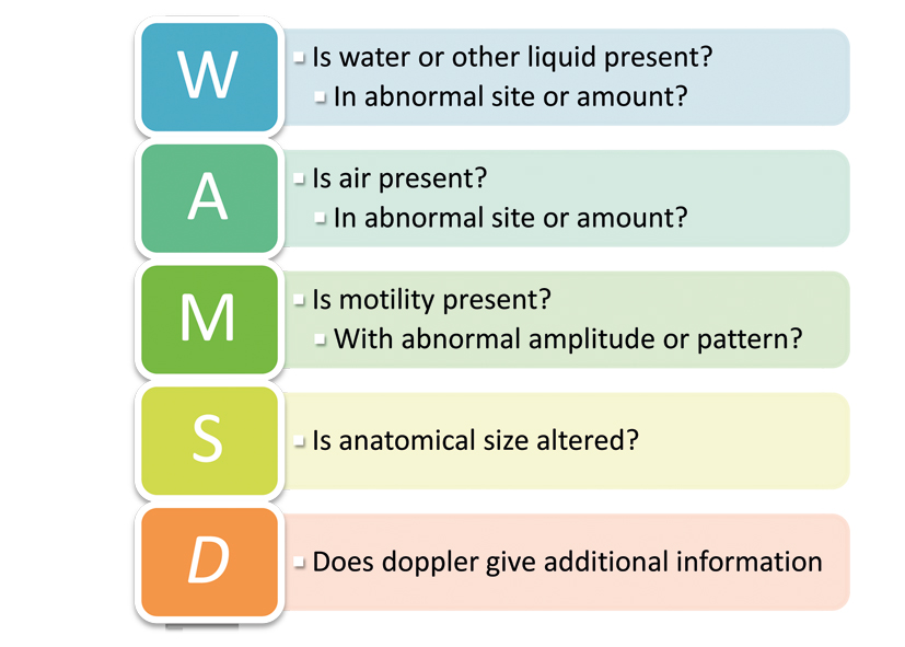
Figure 1. The WAMS-D Approach Algorithm
In the WAMS-D approach, we must begin by asking: Is there liquid (water, blood, urine or other) in our image and, if so, is the position and quantity of such liquid abnormal? Then: Is an air collection hampering ultrasound transmission? Are movements of anatomical parts absent or altered? Are anatomical dimensions altered?
Critical care WAMS ultrasonography can give quick answers in most cases. Doppler gives extra functional information in particular circumstances.
WAMS-D in a Topographic Approach
The following tables, without claiming to be exhaustive, illustrate some of the possible WAMS-D approach applications grouped per anatomical district, as proposed by various authors.
Head
In the head region, precious information can be derived through direct visualisation of the optic nerve and its vascularisation (Rajajee et al. 2011b). Transcranial Doppler, born as a highspecialisation exam, is now increasingly being performed at the bedside by adequately trained intensivists (Munoz- Sanchez et al. 2012).
Table 1. WAMS-D Ultrasonography of the Head
| W | A | M | S | D | |
| Optic nerve | Increased diameter in raised intracranial pressure (Rajajee et al. 2011b) | Monitoring of cerebral hamodynamics (Nenekidis et al. 2011) | |||
| Cerebral arteries | Evaluation of cerebrovascular spasm in haemorrhage (Aaslid et al. 1984) | ||||
| Brain death | Diagnosis of cerebral circulatory arrest (Ducrocq et al. 1998) |
Thorax
Concerning the thoracic region, many
critical cardiac conditions can be rapidly identified by the
intensivist, preceding or, in some cases, substituting the conventional
echocardiography performed by the cardiologist (Vignon 2005).
Table 2. WAMS-D Ultrasonography of the Chest
| W | A | M | S | D | |
| Pleura | Precise evaluation of pleural effusion or empyema (Maslove et al. 2011) | Indirect signs of pneumothorax (Stefanidis et al. 2011) Post-procedural pneumothorax (Vezzani et al. 2011) | Seashore sign in healthy lung (Stefanidis et al. 2011) | ||
| Lung and airways | B-Lines as sign of EVLW (Jambrik et al. 2010) | Assessment of PEEP-induced lung recruitment (Bouhemad et al. 2011) Differential diagnosis of non-pulmonary oedema (Copetti et al. 2008) | Assessment of tracheal trauma (Moriwaki et al. 2006) | ||
| Vessels | Vena cava, vascular filling (De Vecchis et al. 2012) | ||||
| Heart and pericardium | Prompt detection of tamponade (Nagdev and Stone 2011) | Early detection of pneumopericardium (Howlett and Chua 2011) and pneumoperitoneum (Russo and Giangregorio 2012) | Right ventricular strain in pulmonary embolism (Stergiopoulos et al. 2011) | Monitoring volumes (Subramaniam and Talmor 2007) Estimate work of breathing (Vivier et al. 2012) | Focus-oriented echocardiography (Vignon et al. 2007) |
| Diaphragm | Study of diaphgramatic dysfunction (Lerolle et al. 2009) |
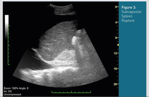
Abdomen
Abdominal CCU has various applications in the study of the hepatobiliary system, pancreas, spleen and genito-urinary system (Wang and Chen 2007). Important data can be obtained from investigation of abdominal vessels: evaluation of volume (Carr et al. 2007) and cases of thrombosis, stenosis or rupture of aneurysms. Protocols have been proposed, reviewed, criticised and refined for rapid assessment in polytrauma patients.
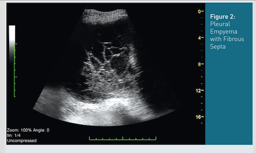
Table 3. WAMS-D Ultrasonography of the Abdomen
| W | A | M | S | D | |
| Abdomen | Free liquid in peritoneum (Tayal et al. 2004) | Perforation (Chen et al. 2002) | Study of biliary tree (Horrow 2010) | Aortic aneurysms, mesenteric infraction (Danse et al. 2009) | |
| Kidney | Hydronephrosis (Barozzi et al. 2007) | Cortical thickness (Barozzi et al. 2007) | Resistance index as shock predictor (Corradi et al. 2011) |
Limbs
In addition to guiding vascular
access, CCU of the limbs can rapidly recognise clots in patients with
suspected deep vein thrombosis, and identify gas presence in cases of
anaerobic soft tissue infections (Jaovisidha et al. 2012).Table 4. WAMS-D Ultrasonography of the Limbs
| W | A | M | S | D | |
| Lower limb | | Soft tissue infection (Jaovisidha et al., 2012) | Deep vein thrombosis (Kory et al., 2011) |
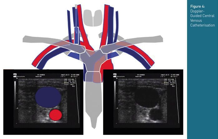
CCU in Invasive Manoeuvres
In addition to the aforementioned diagnostic possibilities, CCU is consolidating its role in assisting the intensivist in the execution of invasive manoeuvres. Some such techniques are widely implemented, like ultrasound-guidance for placement of central venous catheters (Brusasco et al. 2009), execution of pericardiocentesis (Matthew Fields et al. 2012), thoracentesis (Patel et al. 2012) and paracentesis (Nicolaou et al., 2007). Less common applications of CCU include the confirmation of correct endotracheal tube placement (Hsieh et al. 2004), postpyloric feeding tube placement (Hernandez-Socorro et al. 1996) and ultrasound-guided percutaneous dilatational tracheostomy (Rajajee et al. 2011a).
Concluding Remarks
Ultrasonography has countless applications in the intensive care unit. The fact that basic concepts are simple does not mean that the technique is necessarily easy. Primarily, we think attention should be paid on continuous training: the ultrasound machine should be taken out of the closet and each intensivist, from the student to the director, should have the opportunity to train on a daily basis.
It is the authors’ opinion that excessive subjectivity in image interpretation is a major limitation at the moment; therefore, we believe that maximum efforts should be made to develop and validate a quantitative approach to ultrasonography, to reduce effects from operator inexperience, to increase resolution and to obtain a reproducible datum.
References:
Barozzi, L., Valentino, M., Santoro, A., Mancini, E. & Pavlica, P. (2007) "Renal ultrasonography in critically ill patients", Critical care medicine, 35 (5 Suppl) S198-205.
Beaulieu, Y. & Marik, P. E. (2005a) "Bedside ultrasonography in the ICU: part 1", Chest, 128 (2) 881-95.
Beaulieu, Y. & Marik, P. E. (2005b) "Bedside ultrasonography in the ICU: part 2", Chest, 128 (3) 1766-81.
Bouhemad, B., Brisson, H., Le-Guen, M., Arbelot, C., Lu, Q. & Rouby, J. J. (2011) "Bedside ultrasound assessment of positive end-expiratory pressure-induced lung recruitment", American journal of respiratory and critical care medicine, 183 (3) 341-7.
Bouhemad, B., Zhang, M., Lu, Q. & Rouby, J. J. (2007) "Clinical review: Bedside lung ultrasound in critical care practice", Critical care, 11 (1) 205.
Brusasco, C., Corradi, F., Zattoni, P. L., Launo, C., Leykin, Y. & Palermo, S. (2009) "Ultrasound-guided central venous cannulation in bariatric patients", Obesity surgery, 19 (10) 1365-70.
Carr, B. G., Dean, A. J., Everett, W. W., Ku, B. S., Mark, D. G., Okusanya, O., Horan, A. D. & Gracias, V. H. (2007) "Intensivist bedside ultrasound (INBU) for volume assessment in the intensive care unit: a pilot study", The Journal of trauma, 63 (3) 495-500; discussion 500-2.
Chen, S. C., Yen, Z. S., Wang, H. P., Lin, F. Y., Hsu, C. Y. & Chen, W. J. (2002) "Ultrasonography is superior to plain radiography in the diagnosis of pneumoperitoneum", The British journal of surgery, 89 (3) 351-4.
Copetti, R., Soldati, G. & Copetti, P. (2008) "Chest sonography: a useful tool to differentiate acute cardiogenic pulmonary edema from acute respiratory distress syndrome", Cardiovascular ultrasound, 6 16.
Corradi, F., Brusasco, C., Vezzani, A., Palermo, S., Altomonte, F., Moscatelli, P. & Pelosi, P. (2011) "Hemorrhagic shock in polytrauma patients: early detection with renal Doppler resistive index measurements", Radiology, 260 (1) 112-8.
Danse, E. M., Kartheuser, A., Paterson, H. M. & Laterre, P. F. (2009) "Color Doppler sonography of small bowel wall changes in 21 consecutive cases of acute mesenteric ischemia", JBR-BTR : organe de la Societe royale belge de radiologie, 92 (4) 202-6.
De Vecchis, R., Ariano, C., Fusco, A., Ciccarelli, A., Cioppa, C., Giasi, A., Esposito, C. & Cantatrione, S. (2012) "Ultrasound evaluation of the inferior vena cava collapsibility index in congestive heart failure patients treated with intravenous diuretics: new insights about its relationship with renal function: An observational study", Anadolu kardiyoloji dergisi : AKD = the Anatolian journal of cardiology.
Ducrocq, X., Braun, M., Debouverie, M., Junges, C., Hummer, M. & Vespignani, H. (1998) "Brain death and transcranial Doppler: experience in 130 cases of brain dead patients", Journal of the neurological sciences, 160 (1) 41-6.
Gunst, M., Sperry, J., Ghaemmaghami, V., O'keeffe, T., Friese, R. & Frankel, H. (2008) "Bedside echocardiographic assessment for trauma/critical care: the BEAT exam", Journal of the American College of Surgeons, 207 (3) e1-3.
Harding, U., Goeters, C. & Schmidt, C. (2011) "Applications of ultrasound in the intensive care unit", Anasthesiologie, Intensivmedizin, Notfallmedizin, Schmerztherapie : AINS, 46 (3) 190-200; quiz 201.
Hernandez-Socorro, C. R., Marin, J., Ruiz-Santana, S., Santana, L. & Manzano, J. L. (1996) "Bedside sonographic-guided versus blind nasoenteric feeding tube placement in critically ill patients", Critical care medicine, 24 (10) 1690-4.
Hind, D., Calvert, N., Mcwilliams, R., Davidson, A., Paisley, S., Beverley, C. & Thomas, S. (2003) "Ultrasonic locating devices for central venous cannulation: meta-analysis", BMJ, 327 (7411) 361.
Horrow, M. M. (2010) "Ultrasound of the extrahepatic bile duct: issues of size", Ultrasound quarterly, 26 (2) 67-74.
Howlett, P. J. & Chua, T. P. (2011) "Pneumopericardium: diagnosing a rare entity with bedside imaging", Emergency medicine journal : EMJ, 28 (12) 1082.
Hsieh, K. S., Lee, C. L., Lin, C. C., Huang, T. C., Weng, K. P. & Lu, W. H. (2004) "Secondary confirmation of endotracheal tube position by ultrasound image", Critical care medicine, 32 (9 Suppl) S374-7.
Jambrik, Z., Gargani, L., Adamicza, A., Kaszaki, J., Varga, A., Forster, T., Boros, M. & Picano, E. (2010) "B-lines quantify the lung water content: a lung ultrasound versus lung gravimetry study in acute lung injury", Ultrasound in medicine & biology, 36 (12) 2004-10.
Jambrik, Z., Monti, S., Coppola, V., Agricola, E., Mottola, G., Miniati, M. & Picano, E. (2004) "Usefulness of ultrasound lung comets as a nonradiologic sign of extravascular lung water", The American journal of cardiology, 93 (10) 1265-70.
Jaovisidha, S., Leerodjanaprapa, P., Chitrapazt, N., Nartthanarung, A., Subhadrabandhu, T. & Siriwongpairat, P. (2012) "Emergency ultrasonography in patients with clinically suspected soft tissue infection of the legs", Singapore medical journal, 53 (4) 277-82.
Karabinis, A., Fragou, M. & Karakitsos, D. (2010) "Whole-body ultrasound in the intensive care unit: a new role for an aged technique", Journal of critical care, 25 (3) 509-13.
Kendall, J. L., Blaivas, M., Hoffenberg, S. & Fox, J. C. (2004) "History of emergency ultrasound", Journal of ultrasound in medicine : official journal of the American Institute of Ultrasound in Medicine, 23 (8) 1130-3; author reply 1133-5.
Kendall, J. L., Hoffenberg, S. R. & Smith, R. S. (2007) "History of emergency and critical care ultrasound: the evolution of a new imaging paradigm", Critical care medicine, 35 (5 Suppl) S126-30.
Kory, P. D., Pellecchia, C. M., Shiloh, A. L., Mayo, P. H., Dibello, C. & Koenig, S. (2011) "Accuracy of ultrasonography performed by critical care physicians for the diagnosis of DVT", Chest, 139 (3) 538-42.
Lerolle, N., Guerot, E., Dimassi, S., Zegdi, R., Faisy, C., Fagon, J. Y. & Diehl, J. L. (2009) "Ultrasonographic diagnostic criterion for severe diaphragmatic dysfunction after cardiac surgery", Chest, 135 (2) 401-7.
Luecke, T., Corradi, F. & Pelosi, P. (2012) "Lung imaging for titration of mechanical ventilation", Current opinion in anaesthesiology, 25 (2) 131-40.
Maslove, D. M., Chen, B. T., Wang, H. & Kuschner, W. G. (2011) "The Diagnosis and Management of Pleural Effusions in the ICU", Journal of intensive care medicine.
Matthew Fields, J., Paziana, K., Vuljaj, N., Fischer, J. & Ku, B. S. (2012) "Emergency Ultrasound-guided Pericardiocentesis Using a High-frequency Linear Array Transducer to Improve Needle Tip Visualization", Academic emergency medicine : official journal of the Society for Academic Emergency Medicine.
Moriwaki, Y., Sugiyama, M., Fujita, S., Toyoda, H., Kosuge, T., Yamamoto, T., Amano, S., Matsuzaki, S., Shimoyama, T., Tahara, Y., Iwashita, M., Fukuyama, H. & Suzuki, N. (2006) "Application of ultrasonography for blunt laryngo-cervical-tracheal injury", The Journal of trauma, 61 (5) 1156-61.
Munoz-Sanchez, M. A., Murillo-Cabezas, F., Egea-Guerrero, J. J., Gascon-Castillo, M. L., Cancela, P., Amaya-Villar, R., Rincon-Ferrari, M. D., Flores-Cordero, J. M., Cayuela, A. & Garcia-Alfaro, C. (2012) "Emergency transcranial doppler ultrasound: predictive value for the development of symptomatic vasospasm in spontaneous subarachnoid hemorrhage in patients in good neurological condition", Medicina intensiva / Sociedad Espanola de Medicina Intensiva y Unidades Coronarias.
Nagdev, A. & Stone, M. B. (2011) "Point-of-care ultrasound evaluation of pericardial effusions: does this patient have cardiac tamponade?", Resuscitation, 82 (6) 671-3.
Nenekidis, I., Geiser, M., Riva, C., Pournaras, C., Tsironi, E., Vretzakis, G., Mitilis, V. & Tsilimingas, N. (2011) "Blood flow measurements within optic nerve head during on-pump cardiovascular operations. A window to the brain?", Interactive cardiovascular and thoracic surgery, 12 (5) 718-22.
Nicolaou, S., Talsky, A., Khashoggi, K. & Venu, V. (2007) "Ultrasound-guided interventional radiology in critical care", Critical care medicine, 35 (5 Suppl) S186-97.
Patel, P. A., Ernst, F. R. & Gunnarsson, C. L. (2012) "Ultrasonography guidance reduces complications and costs associated with thoracentesis procedures", Journal of clinical ultrasound : JCU, 40 (3) 135-41.
Rajajee, V., Fletcher, J. J., Rochlen, L. R. & Jacobs, T. L. (2011a) "Real-time ultrasound-guided percutaneous dilatational tracheostomy: a feasibility study", Critical care, 15 (1) R67.
Rajajee, V., Vanaman, M., Fletcher, J. J. & Jacobs, T. L. (2011b) "Optic nerve ultrasound for the detection of raised intracranial pressure", Neurocritical care, 15 (3) 506-15.
Russo, A. & Giangregorio, C. (2012) "[Pneumomediastinum: an extremely rare affection. A case of deferred diagnosis. Literature review and the role of thoracic US in urgency]", Annali italiani di chirurgia, 83 (1) 13-9.
Stefanidis, K., Dimopoulos, S. & Nanas, S. (2011) "Basic principles and current applications of lung ultrasonography in the intensive care unit", Respirology, 16 (2) 249-56.
Stergiopoulos, K., Bahrainy, S., Strachan, P. & Kort, S. (2011) "Right ventricular strain rate predicts clinical outcomes in patients with acute pulmonary embolism", Acute cardiac care, 13 (3) 181-8.
Subramaniam, B. & Talmor, D. (2007) "Echocardiography for management of hypotension in the intensive care unit", Critical care medicine, 35 (8 Suppl) S401-7.
Sustic, A., Kovac, D., Zgaljardic, Z., Zupan, Z. & Krstulovic, B. (2000) "Ultrasound-guided percutaneous dilatational tracheostomy: a safe method to avoid cranial misplacement of the tracheostomy tube", Intensive care medicine, 26 (9) 1379-81.
Tayal, V. S., Beatty, M. A., Marx, J. A., Tomaszewski, C. A. & Thomason, M. H. (2004) "FAST (focused assessment with sonography in trauma) accurate for cardiac and intraperitoneal injury in penetrating anterior chest trauma", Journal of ultrasound in medicine : official journal of the American Institute of Ultrasound in Medicine, 23 (4) 467-72.
Vezzani, A., Brusasco, C., Palermo, S., Launo, C., Mergoni, M. & Corradi, F. (2010) "Ultrasound localization of central vein catheter and detection of postprocedural pneumothorax: an alternative to chest radiography", Critical care medicine, 38 (2) 533-8.
Vignon, P. (2005) "Hemodynamic assessment of critically ill patients using echocardiography Doppler", Current opinion in critical care, 11 (3) 227-34.
Vignon, P., Dugard, A., Abraham, J., Belcour, D., Gondran, G., Pepino, F., Marin, B., Francois, B. & Gastinne, H. (2007) "Focused training for goal-oriented hand-held echocardiography performed by noncardiologist residents in the intensive care unit", Intensive care medicine, 33 (10) 1795-9.
Vivier, E., Mekontso Dessap, A., Dimassi, S., Vargas, F., Lyazidi, A., Thille, A. W. & Brochard, L. (2012) "Diaphragm ultrasonography to estimate the work of breathing during non-invasive ventilation", Intensive care medicine, 38 (5) 796-803.
Wang, H. P. & Chen, S. C. (2007) "Upper abdominal ultrasound in the critically ill", Critical care medicine, 35 (5 Suppl) S208-15.