A research team at the Victor Chang Cardiac Research Institute in Australia has created an integrated map of heart cells that unlocks cardiac fibrosis, a leading cause of heart failure. The study is published in Science Advances.
This breakthrough paves the way for the development of targeted drugs to prevent scarring that occurs after a heart attack. During and after a heart attack, the heart muscle is damaged, resulting in scar tissue formation. Unlike healthy heart muscle, scar tissue lacks elasticity and contractility, leading to permanent damage and impaired blood pumping, eventually causing heart failure.
This study improves the understanding of cardiac fibrosis, which is prevalent in virtually all heart diseases, including those caused by high blood pressure.
Significant investment has been made in finding new drugs to control cardiac fibrosis, but these efforts have largely failed. There is a critical need to develop novel treatments that could halt or even reverse cardiac fibrosis, benefiting millions, the researchers note.
Fibrosis is a natural part of the body’s healing process. However, in the heart, if the disease triggers are unresolved, it can lead to excessive scarring, severely impairing heart function and becoming a major cause of heart failure.
For the first time, using revolutionary technology to analyse gene expression in single cells, the team has mapped the progressive cell states involved in cardiac fibrosis and how these cells evolve day by day. They analysed RNA signatures from 100,000 single cells, focusing on those involved in fibrosis and integrating data from various pioneering studies on different heart disease states. This allowed them to produce an integrated cellular map of a mouse model heart, identifying cells and pathways involved in fibrosis.
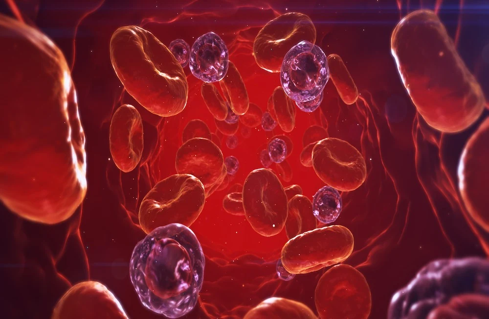
They identified resting cells, activated cells, an inflammatory population, a progenitor pool, dividing cells, and specialised cells called myofibroblasts and matrifibrocytes. The study discovered that myofibroblasts, major drivers of scarring absent in healthy hearts, begin forming three days after a heart attack in a mouse model, peaking at day five, and then transitioning to matrifibrocytes, which may prevent the scar from resolving.
The study also explored other heart disease models, such as heart failure induced by high internal blood pressure due to aortic stenosis or hypertension. The researchers found a surprising similarity in fibrosis progression across different heart diseases. Myofibroblasts were abundant early on during hypertension and then resolved into matrifibrocytes, just as they do after a heart attack.
This discovery opens doors to future therapies that target specific cell types or processes in different heart diseases, potentially preventing healthy cells from being permanently damaged.
The study utilised data from both mouse models and human patients. Heart failure can take decades to develop in humans, so the exact cell types and timing of processes in human patients require further detailed exploration.
Persistent high blood pressure can have devastating consequences, but it is treatable. Thus monitoring and controlling high blood pressure promptly is extremely important.
Source: Victor Change Cardiac Research Institute
Image Credit: iStock


