Researchers have tested a new more nuanced form of investigating the subtle clues sent out by the human body during anaesthesia — particularly the cardiovascular signals that can indicate the state of the pain-monitoring autonomous nervous system. The results have proved more reliable than existing methods in monitoring depth of anaesthesia, according to a new study published in the journal Anaesthesia.
The breakthrough could help to provide a new guide for anaesthetists and lead to much quicker recovery times for patients following operations as greater optimisation of dosage could lead to drugs being significantly reduced.
"The likelihood of waking up during surgery is extremely small but, if it happened, it could be a distressing experience," said Peter McClintock, PhD, a professor of physics at Lancaster University and co-author of the study. "So we are delighted to pave the way to a new tool for gauging depth of anaesthesia."
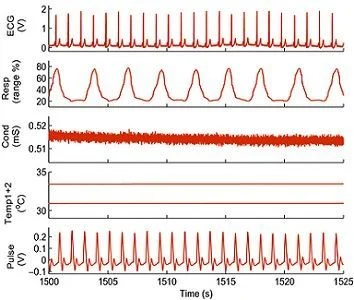
The researchers found that by looking at how key indicators (ie, respiration, ECG, skin temperature, pulse and skin conductivity) interacted with one another, they could better predict whether a patient was awake or anaesthetised. These indicators also helped distinguish between the effects of two commonly-used anaesthetic drugs, propofol and sevoflurane.
The study measured the state of anaesthesia of 27 patients in good health during surgery in the UK and Norway. Readings were taken at a high frequency — several hundred samples per heart beat — for 30 minutes while the patients were awake before their surgery and for up to 30 minutes during general anaesthesia. The signals showed, with a high degree of accuracy, how the patients reacted to the anaesthetics, according to the researchers.
“We have developed new methods to study complex interactions between ever changing processes such as the processes in our heart, lungs and vasculature," said co-author Professor Aneta Stefanovska, also of Lancaster University. "These physiological processes constantly interact with one another but anaesthetic drugs change the level of these interactions. By applying our new methods, in this study we were able to get a very accurate picture of what was going on, leading to the most reliable predictions of the state of anaesthesia obtained from cardiovascular signals to date of closer to 97 percent.”
The collaborative research involved consultant anaesthetists from University Hospitals of Morecambe Bay NHS Trust in North West England.
Professor Andrew Smith, Consultant Anaesthetist at Royal Lancaster Infirmary, who also took part in the study, said: "Whilst it is early days, the prospect of a monitor of anaesthetic depth that relies on measurements of the circulation and respiration is very attractive. I look forward to further collaboration with Lancaster University in the interests of enhancing the care of patients in the NHS locally and nationally in the future.”
Source: Lancaster University
Image credit: Anaesthesia
The breakthrough could help to provide a new guide for anaesthetists and lead to much quicker recovery times for patients following operations as greater optimisation of dosage could lead to drugs being significantly reduced.
"The likelihood of waking up during surgery is extremely small but, if it happened, it could be a distressing experience," said Peter McClintock, PhD, a professor of physics at Lancaster University and co-author of the study. "So we are delighted to pave the way to a new tool for gauging depth of anaesthesia."
The researchers found that by looking at how key indicators (ie, respiration, ECG, skin temperature, pulse and skin conductivity) interacted with one another, they could better predict whether a patient was awake or anaesthetised. These indicators also helped distinguish between the effects of two commonly-used anaesthetic drugs, propofol and sevoflurane.
The study measured the state of anaesthesia of 27 patients in good health during surgery in the UK and Norway. Readings were taken at a high frequency — several hundred samples per heart beat — for 30 minutes while the patients were awake before their surgery and for up to 30 minutes during general anaesthesia. The signals showed, with a high degree of accuracy, how the patients reacted to the anaesthetics, according to the researchers.
“We have developed new methods to study complex interactions between ever changing processes such as the processes in our heart, lungs and vasculature," said co-author Professor Aneta Stefanovska, also of Lancaster University. "These physiological processes constantly interact with one another but anaesthetic drugs change the level of these interactions. By applying our new methods, in this study we were able to get a very accurate picture of what was going on, leading to the most reliable predictions of the state of anaesthesia obtained from cardiovascular signals to date of closer to 97 percent.”
The collaborative research involved consultant anaesthetists from University Hospitals of Morecambe Bay NHS Trust in North West England.
Professor Andrew Smith, Consultant Anaesthetist at Royal Lancaster Infirmary, who also took part in the study, said: "Whilst it is early days, the prospect of a monitor of anaesthetic depth that relies on measurements of the circulation and respiration is very attractive. I look forward to further collaboration with Lancaster University in the interests of enhancing the care of patients in the NHS locally and nationally in the future.”
* * * * * * * * * * *
Top image. Example of a short segment of signals recorded during anaesthesia (from top to bottom): electrical activity of the heart (ECG); respiration as a percentage of the sensor range; skin conductivity; skin temperature from the wrist (upper) and ankle (lower) and piezoelectric pulse.Source: Lancaster University
Image credit: Anaesthesia
References:
Smith AF, McClintock PVE, Stefanovska A et al. (2015) The discriminatory value of cardiorespiratory interactions in distinguishing awake from anaesthetised states: a randomised observational study. Anaesthesia. 9
SEP 2015. DOI: 10.1111/anae.13208
Latest Articles
healthmanagement, anaesthesia, surgery, respiration, cardiovascular, heart beat
Researchers have tested new more nuanced form of investigating the subtle clues sent out by the human body during anaesthesia — particularly the cardiovascular signals that can indicate the state of the pain-monitoring autonomous nervous system.


![Tuberculosis Diagnostics: The Promise of [18F]FDT PET Imaging Tuberculosis Diagnostics: The Promise of [18F]FDT PET Imaging](https://res.cloudinary.com/healthmanagement-org/image/upload/c_thumb,f_auto,fl_lossy,h_184,q_90,w_500/v1721132076/cw/00127782_cw_image_wi_88cc5f34b1423cec414436d2748b40ce.webp)


