Gout is a condition where monosodium urate (MSU) crystals cause granulomatous inflammation, forming tophi in joints and soft tissues, mainly in the lower extremities. It typically manifests as acute monoarticular flares but can present atypically or chronically, mimicking other arthritis types. Dual-energy computed tomography (DECT) effectively detects MSU crystals by exploiting different tissue attenuation at varying X-ray energies. Contrast-enhanced multimodal DECT enhances this capability, aiding in evaluating soft tissue inflammation, erosions, and other lesions. Virtual noncontrast (VNC) imaging, a DECT postprocessing technique, subtracts iodine maps from contrast-enhanced images to create native-like scans. This method aims to improve MSU crystal identification despite challenges posed by high iodine contrast concentrations. The aim of a study recently published in European Radiology Experimental was to investigate MSU detection in VNC images generated from contrast-enhanced DECT datasets.
Phantom-Based Evaluation of VNC Imaging for MSU Crystal Detection in DECT
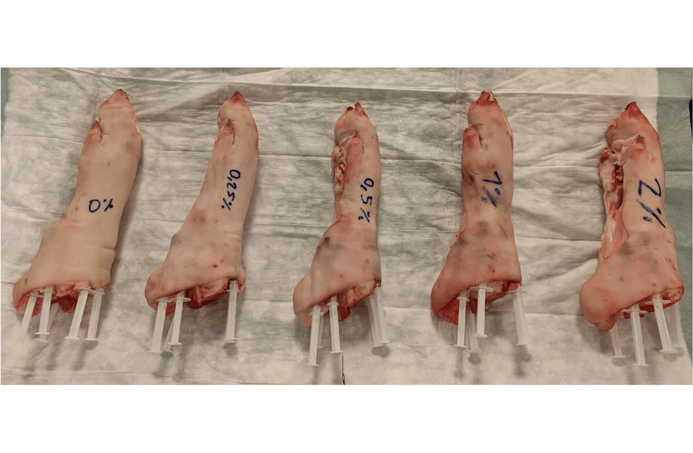
In this study, two phantom models were used to simulate varying concentrations of monosodium urate (MSU) crystals in vivo. The first phantom involved 25 syringes with different mixtures of MSU, ultrasound gel, and iodinated contrast medium (ICM), arranged in a grid immersed in water. The second phantom used porcine forelegs to mimic realistic gouty tophi, with syringes containing MSU placed around the joints. Both phantoms were scanned using a 320-row CT scanner in dual-energy mode with sequential scans at 80 kVp and 135 kVp. Image reconstruction was done with adaptive iterative dose reduction, yielding VNC (virtual noncontrast) images through iodine map subtraction. These VNC images were processed using a two-material decomposition algorithm to detect MSU crystals, with results compared to conventional CT images processed similarly.
Quantitative analysis involved recording MSU volumes and masses detected, while false-positive detections were noted as artifacts. A detection index (DI) was used to enhance result comparability across different MSU concentrations. Statistical analysis, including paired t-tests, was employed to assess the significance of findings between VNC and conventional CT images. The study aimed to validate the efficacy of VNC imaging in detecting MSU crystals under various conditions, providing insights into its potential clinical utility for improving gout diagnosis and management decisions.
VNC imaging effectively mitigated the negative impact of high ICM concentrations on MSU detection
In this phantom study, virtual noncontrast (VNC) imaging was evaluated for its ability to detect monosodium urate (MSU) crystals amidst high concentrations of iodinated contrast medium (ICM), overcoming previous challenges observed with dual-energy computed tomography (DECT). The study employed two-material decomposition analysis to assess MSU volumes and estimated masses, using a detection index (DI) to compare results across different ICM concentrations.
The findings indicated that VNC imaging effectively mitigated the negative impact of high ICM concentrations on MSU detection, which had been reported in previous research. Specifically, VNC postprocessing successfully reduced the effective atomic number (Zeff) in voxels containing MSU crystals, thereby enhancing their characterization despite the presence of iodine. Conversely, samples with low or no ICM showed reduced sensitivity in VNC imaging, attributed to the absence of iodine-enhanced attenuation effects.
The study highlighted the technical nuances of DECT, where iodinated contrast can influence voxel density and Zeff, potentially affecting MSU detection thresholds. It also noted limitations in the current VNC methodology, particularly in accurately differentiating between MSU and other tissues like calcium deposits in joints. Recommendations included using both VNC and conventional CT imaging approaches when assessing gouty tophi to ensure comprehensive diagnostic evaluation.
Despite these insights, the study acknowledged limitations such as potential syringe content distribution variations and the need for further research in clinical settings to validate these findings. Overall, the study underscores the potential of VNC imaging in improving the diagnostic utility of contrast-enhanced DECT for gout detection, albeit with considerations for optimizing postprocessing techniques and standardizing protocols across different imaging systems.
Source & Image Credit: European Radiology Experimental


