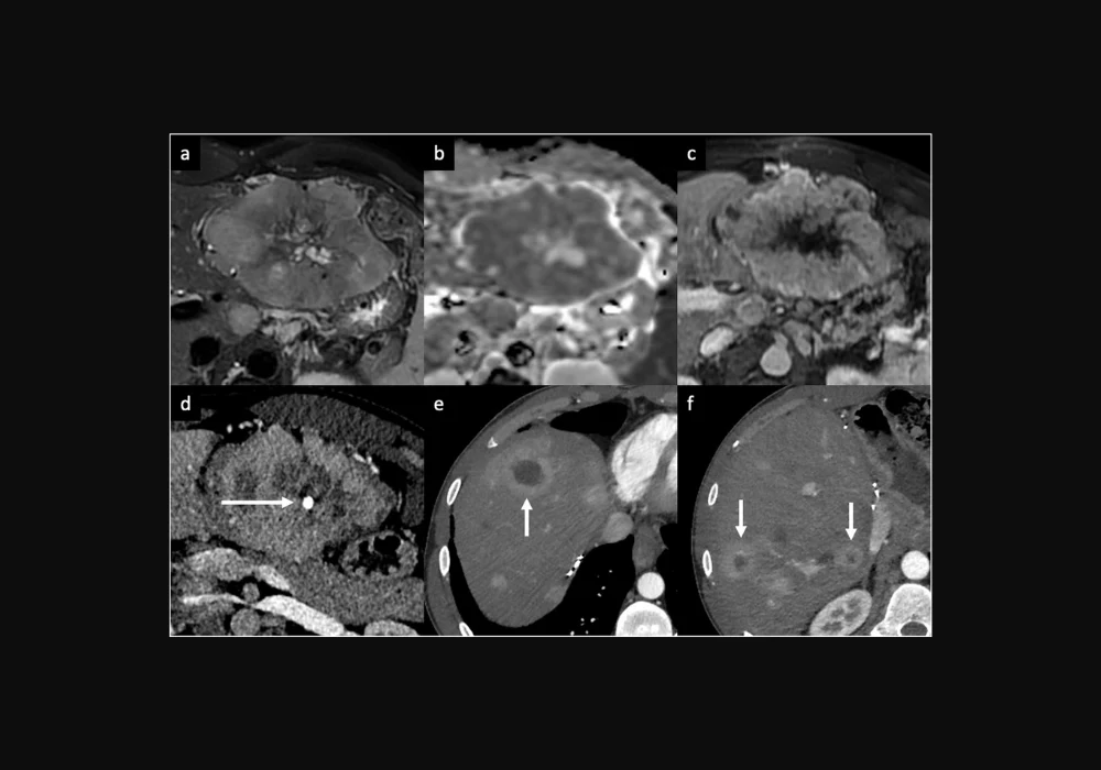
Peripheral arterial enhancement (PAE) of focal liver lesions represents a crucial imaging feature observed in the arterial phase (AP) of contrast-enhanced CT and MRI. This feature encompasses various patterns, such as peripheral nodular discontinuous enhancement, rim arterial phase hyperenhancement (rim APHE), and corona enhancement. Identifying and differentiating these patterns is vital to diagnosing benign and malignant liver lesions. This article aims to provide a detailed review of the broad spectrum of focal liver lesions exhibiting rim APHE, discussing their imaging characteristics, clinical significance, and differentiation strategies.
Benign Lesions with Peripheral Arterial Enhancement
♦ Vascular Lesions: Sclerosed Hemangiomas
Sclerosed hemangiomas are a rare end stage of hemangioma involution, characterised by extensive fibrosis, narrowing or obliteration of vascular spaces, and hemorrhage or sclerosis. These lesions often occur in cirrhotic livers and can exhibit capsular retraction and fibrotic changes over time. On contrast-enhanced CT, sclerosed hemangiomas may display a rim APHE that persists into the portal venous phase (PVP), with irregular delayed enhancement regions. MRI characteristics include variable signal intensity (SI) on T2-weighted imaging (WI), low or heterogeneous SI on T1-WI, and high SI on T2-WI. The presence of T2-shine-through on the apparent diffusion coefficient (ADC) map and high SI on T2-WI are key radiological features. Challenging cases may benefit from ultra-delayed phase acquisitions to observe centripetal enhancement and contrast retention.
♦ Infectious Lesions: Abscesses
Liver abscesses, resulting from hematogenous dissemination of infections or direct bacterial introduction, present distinct imaging features. Clinically, abscesses manifest with fever, abdominal pain, and elevated inflammatory markers. On contrast-enhanced CT, pyogenic abscesses appear as well-defined hypoattenuating lesions with a layered wall and early inner wall rim APHE that persists in the delayed phases, often surrounded by transient perfusion disorders. MRI shows central low SI on T1-WI, high SI on T2-WI, and the double target sign on diffusion-weighted imaging (DWI). Differentiating pyogenic from amebic abscesses can be challenging, with solitary abscesses more likely to be amebic. Occasionally, abscesses may mimic hepatic tumours like intrahepatic cholangiocarcinoma (iCCA), necessitating aspiration or biopsy for confirmation.
♦ Inflammatory Lesions: Granulomatous Diseases
Granulomatous hepatitis, often associated with sarcoidosis, tuberculosis, and histoplasmosis, may occasionally present with multiple small hypoattenuating lesions showing subtle rim APHE on imaging. MRI reveals hypointense lesions on T1-WI and hypo-to-isointense on T2-WI. These nonspecific findings often require percutaneous liver biopsy for definitive diagnosis. Solitary necrotic nodules, another inflammatory lesion, appear as small solitary nodules with rim APHE and thin delayed rim enhancement early in development, evolving to show calcifications and lack of enhancement as they mature.
Malignant Lesions with Peripheral Arterial Enhancement
♦ Intrahepatic Cholangiocarcinoma (iCCA)
iCCA, the most common primary non-HCC malignancy in the liver, typically presents as a large lesion with irregular lobulated margins and rim APHE. This enhancement pattern reflects the tumour's histology, with viable cells at the periphery and a central desmoplastic stroma. MRI features include low-to-moderate SI on T2-WI and low SI on T1-WI, with a targetoid appearance on high b-value DWI and hepatobiliary phase (HBP) of gadoxetate disodium-enhanced MRI (Gd-EOB-MRI). iCCA often shows additional findings like capsular retraction, bile duct dilatation, and vascular encasement, distinguishing it from other liver lesions. Serum tumour markers such as CA 19-9 aid in the diagnosis, but histopathologic confirmation remains essential.
♦ Hepatocellular Carcinoma (HCC)
HCC is the leading cause of cancer-related mortality in chronic liver disease patients, commonly associated with chronic viral hepatitis and metabolic dysfunction. Typical imaging features include non-rim APHE and non-peripheral washout on PVP or delayed phases. However, 5.6-15.7% of HCCs may display an atypical peripheral rim APHE, often seen in fibrotic, poorly differentiated, or sarcomatoid subtypes. The presence of ancillary features like non-enhancing capsules, mosaic architecture, and intralesional fat can guide the diagnosis. LI-RADS criteria and patient risk status are crucial in differentiating HCC from other liver malignancies.
♦ Metastatic Lesions
Liver metastases are more common than primary liver cancers, often exhibiting rim APHE on dynamic imaging. Hypoenhancing metastases from adenocarcinomas and hyper enhancing ones from neuroendocrine tumours or renal cell carcinoma shows early rim APHE while hyper enhancing ones exhibit delayed rim enhancement. MRI, particularly with hepatobiliary contrast agents, offers higher sensitivity for detecting small liver metastases. Recognising characteristic imaging features and patient history is essential for accurate diagnosis and treatment planning.
Peripheral arterial enhancement of focal liver lesions encompasses a wide range of benign and malignant entities, each with distinct imaging characteristics. Understanding these patterns and integrating clinical, laboratory, and imaging findings are crucial for accurate diagnosis and management. Histopathological examination remains the gold standard for definitive diagnosis in ambiguous cases. Advances in imaging techniques and contrast agents continue to enhance the ability to differentiate between various liver lesions, ultimately improving patient outcomes.
Source & Image Credit: Insights into Imaging


