During 2019, a major area of interest in cardiology was multimodality imaging and the application of artificial intelligence to imaging techniques. While conventional imaging still remains important for the assessment of cardiac structure and function, advanced echocardiography with strain imaging techniques, tissue characterisation with cardiovascular magnetic resonance (CMR), and the assessment of biological processes with nuclear imaging techniques have helped clinicians implement early intervention strategies to either prevent or halt the progression of cardiovascular disease.
You might also like: Nuclear Cardiology: Molecular Insights into the Heart
Overall, in 2019, the application of AI and machine learning to cardiac imaging has been a major novelty. In addition, there were advances in non-invasive cardiac imaging that also helped improve diagnosis and treatment of patients.
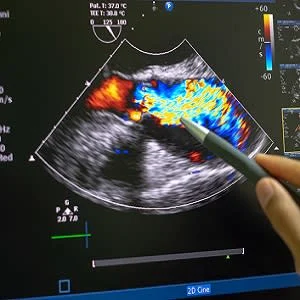
The Copenhagen City Heart Study evaluated the association between left ventricular filling pressures measured on tissue Doppler imaging, and speckle tracking echocardiography with the occurrence of cardiovascular death, hospital admission for incident heart failure or myocardial infarction. Findings showed that speckle tracking echocardiography was independently associated with the endpoint. It also provided incremental prognostic value over the SCORE risk chart that evaluates the risk of cardiovascular morbidity and mortality.
Echocardiography is also the imaging technique of choice to assess valvular heart disease. Also, the use of transthoracic focused cardiac ultrasound (FoCUS) has gained popularity in emergency departments and intensive care units. In addition, elevated carotid artery wave intensity, as measured on Duplex Doppler ultrasound, was found to be independently associated with faster cognitive decline in the Whitehall II study. These findings highlight the relevance and importance of ultrasound imaging outside the heart.
In recent years, cardiovascular magnetic resonance has made a significant contribution to the diagnosis and management of cardiomyopathies. Similarly, computed tomography has also now evolved into a one-stop-shop imaging tool providing valuable diagnostic and prognostic information in patients with suspected or known coronary artery disease. Coronary artery calcium (CAC) score on CT was also increasingly used to guide statin therapy.
There is no doubt that invasive and non-invasive cardiovascular imaging is developing at a rapid pace. Today, clinicians have access to different imaging modalities, which are now even more effective with fusion imaging as well as the introduction of AI and machine learning. In particular, machine learning has been rapidly adopted and allows automated analysis of electrocardiograms, echocardiography, nuclear perfusion imaging and CCTA.
Finally, deep learning has also been applied quite effectively to analyse echocardiographic data, including view identification, image segmentation, quantification of structure and function and disease detection.
Overall, imaging in cardiology saw significant advancement in 2019 and this trend is expected to continue in 2020 as we will likely see a consistent application of artificial intelligence, machine learning and deep learning in healthcare overall, and imaging specifically.
Source: European Heart Journal
Image Credit: iStock
References:
Pennell D et al. (2020) The year in cardiology: imaging: The year in cardiology 2019. European Heart Journal, ehz930.
Latest Articles
CT, CCTA, nuclear imaging, echocardiography, CMR
One of the main novelties of 2019 was the application of artificial intelligence and machine learning to cardiac imaging, as well as advances in non-invasive cardiac imaging.


