


Materials scientists, led by Bert Müller (Biomaterials Science Center at University of Basel) have developed a new way to visualise constricted and calcified blood vessels with micrometer precision.
The “NO-stress” project combined hard x-ray tomography and established histology methods to visualise the vessels constricted by atherosclerosis. Atherosclerotic plaque shows strong contrast in x-rays, and it has been very difficult to identify soft tissues in the direct neighbourhood of calcifications using x-rays.
The data about the morphology of the constricted vessels is used to simulate blood flow and find out related shear stresses. Shear stress is increased at the constrictions, and is the basis for the development of specialised nano-containers for the targeted and local delivery of vasodilation drugs
The new method is also suitable for three-dimensional characterisation of any other combination of strongly and weakly x-ray absorbing species, such as cartilage and bone. It takes advantage of conventional x-ray absorption and of x-ray phase contrast measurements, which are accessible via grating interferometry. As phase contrast is less dependent on the atomic number of the constituents than absorption contrast, soft tissues in the vicinity of hard tissues can be more easily visualised.
The authors show that strongly calcified arteries are thoroughly characterised by the combination of the non-destructive tomography measurements in x-ray absorption and phase contrast modes, together with established histology techniques.
The project “NO-stress” is funded within the National Research Programme NRP 62 “Smart Materials” by the Swiss National Science Foundation.
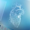
Image caption:
Conventional micro-tomography using intense x-rays allows for the visualisation of plaque (white) and muscle tissue (black). Here, the blood vessel walls are invisible. More sophisticated x-ray computed tomography techniques enable the identification of the vessel walls and other soft tissues. The images show experimental data acquired at the beam-line BW2 (HASYLAB at DESY, Germany)
Source: AlphaGalileo
Image credit: Biomaterials Science Center, University of Basel, Switzerland.
References:
Reference
Holme MN, Schulz G, Deyhle H et al. (2014) Complementary X-ray tomography techniques for histology-validated three-dimensional imaging of soft and hard human tissues. Nature Protocols, 9: 1401-15. doi:10.1038/nprot.2014.091
Latest Articles
atherosclerosis, medical imaging
Materials scientists, led by Bert Müller (Biomaterials Science Center at University of Basel) have developed a new way to visualise constricted and calcif...