Researchers at the Oxford University have developed a technique that could improve heart scans for patients and could provide more information about the heart as compared to traditional scans. The new technique will not require any injections and will be much safer and faster.
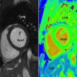
The research team, based at the Oxford Centre for Clinical Magnetic Resonance Research (OCMR), used a property of hydrogen atoms and created a pixel-by-pixel map of the heart. The map, called T1, allows examination of both healthy and diseased heart tissue in much greater detail than traditional scans. T1 is the time constant that describes the speed with which atoms return to a normal thermodynamic state after being affected by radio waves and magnetic fields.
Traditional scans that use MRI require patients to be injected with two substances - adenosine and gadolinium. However, the research team at Oxford used T1 mapping to see if that would give more clearer and more clinically useful results. When doctors use traditional scans, they use shades of light and dark to judge the condition of the heart. But with T1 maps, the results are less subjective since it provides colour-coded numbers.
These scans would also be more useful in patients with severe kidney failure as they cannot clear Gadolinium and are often unable to benefit from the regular MRI scan. T1 mapping enables the doctors to see the presence of more water, which is often the case in heart conditions. By visualising T1 values across the heart, it would be possible to determine the precise location of the problem.
The new technique is very fast and takes only three minutes to map the whole thing. The values that it gives turn into a colour map providing doctors an image that is quicker to understand and less subjective.
Dr Stefan Piechnik who developed the specific T1 mapping technique at Oxford, explains, 'T1 mapping allows us to look in finer detail at the heart in a non-invasive way, which has not been possible before. We can now get results without Gadolinium, meaning we have a technique that is safer and quicker and can be used with more people. The results are also less dependent on interpreting the images -- medics have something based on hard numbers.'
There is no doubt that this research holds great potential. The ability to provide pixel by pixel detail can help identify unhealthy tissues wherever they appear. The technique is already being applied in studies with the goal that it would be available for clinical use within the next five years.
Source: University of Oxford
Image Credit: Dr Alexander Liu/ OCMR/ University of Oxford

