The first recommendations on multimodality imaging assessment of prosthetic heart valves have been published in European Heart Journal. The paper outlines how multimodality imaging should be used to detect and diagnose prosthetic heart valve complication
Prosthetic heart valves are often inserted in patients who have disease of their native valves and are symptomatic. Valvular heart disease is quite common and accounts for about 3-6% of the cases of heart disease in people over the age of 65. In fact in developing countries, valvular heart disease due to rheumatic fever accounts for the majority of heart disease.
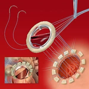
When the heart valves fail to work, they can be replaced with a biological or a mechanical prosthesis. It is estimated that by the year 2050, some 850,000 prosthetic heart valves will have been implanted in people living in western nations. However, like any mechanical device, heart prosthesis do not last forever. The life span of a heart valve ranges from 10-20 years, depending on the patient's age and the type of valve. When a a heart valve starts to malfunction, the patient may again develop symptoms. When this occurs, it is important to know the cause of the heart valve dysfunction so that the appropriate therapy can be offered. When prosthetic heart valve complications are suspected, the authors recommend:
- First-line imaging with 2D transthoracic echocardiography (TTE)
- 2D and 3D TTE and transoesophageal echocardiography (TOE) for complete evaluation
- Cinefluoroscopy to evaluate disc mobility and valve ring structure
- Cardiac computed tomography (CT) to visualise calcification, degeneration, pannus, thrombus
- Cardiac magnetic resonance imaging (CMR) to assess cardiac and valvular function
- Nuclear imaging, especially when infective endocarditis is suspected.
None of these studies alone is perfect and none can detect mild cases of prosthetic health valve malfunction. Echo is usually the study of choice and non-echo modalities are used if more detailed information is required. To improve the sensitivity of the studies, researchers have now developed a new algorithm which clinicians can follow to help make an earlier diagnosis of prosthetic valve dysfunction and also quantify the dysfunction.
So far there are no long term data on how this algorithm has improved imaging of prosthetic heart valves. It is also important to remember that many of the non-echo modalities for assessing the heart valve are not routinely available in all hospitals and are also prohibitively expensive.
Source: European Heart Journal
Image Credit: Wikimedia Commons


