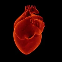
Large cardiac-muscle patches created from human cells have been tested on large animals in a heart attack model, with results showing the patches significantly improved recovery from heart attack injury, according to a study published in the journal Circulation. The results are a step closer to the goal of treating human heart attacks by suturing cardiac-muscle patches over an area of dead heart muscle in order to reduce the pathology that often leads to heart failure.
Researchers created the patches by suspending cardiomyocytes, smooth-muscle cells, and endothelial cells that had been differentiated from human induced-pluripotent stem cells (hiPSCs) in a fibrin scaffold and then culturing the construct on a dynamic (rocking) platform. The research team was led by Jianyi Zhang, MD, PhD, the chair of University of Alabama at Birmingham Biomedical Engineering, a joint department of the UAB School of Medicine and the UAB School of Engineering.
Each patch is 1.57 by 0.79 inches in size and nearly as thick as a dime. Zhang and colleagues found that transplanting two of these patches onto the infarcted area of a pig heart significantly improved function of the heart’s left ventricle, the major pumping chamber. In addition, the patches significantly reduced infarct size, which is the area of dead muscle; heart-muscle wall stress and heart-muscle enlargement; as well as significantly reducing apoptosis, or programmed cell death, in the scar boarder area around the dead heart muscle.
Notably, use of these patches did not induce arrhythmia in the hearts, a serious complication observed in some past biomedical engineering approaches to treating heart attacks.
A key to success of the patches is how they are engineered, the researchers explain. Each patch is a mixture of three cell types — 4 million cardiomyocytes, or heart-muscle cells; 2 million endothelial cells, which are well-known to help cardiomyocytes survive and function in a micro-environment; and 2 million smooth muscle cells, which line blood vessels. Each patch was grown in a three-dimensional fibrin matrix that was rocked back and forth for a week. The cells begin to beat synchronously after one day. The patches resembled native heart-muscle tissue in their physiological and contractile properties.
Previous attempts to use hiPSCs to treat animal models of heart attacks — using an injection of cells or cells grown as a very thin film — have shown very low rates of survival, or engraftment, by the hiPSCs. The present study had a relatively high rate of engraftment, 10.9 percent, four weeks after transplantation, and the transplantation led to improved heart recovery.
Part of the beneficial effects of the patches may occur through the release of tiny blebs called exosomes from cells in the patches. These exosomes, which carry proteins and RNA from one cell to another, are a common cell-to-cell signalling method that is incompletely understood. In tissue culture experiments, the researchers found that exosomes released from the large heart-muscle patches appeared to protect the survival of heart-muscle cells.
Source: University of Alabama at Birmingham
Image Credit: University of Alabama, Birmingham
Latest Articles
heart muscle, heart attacks, cardiac-muscle patches
Large cardiac-muscle patches created from human cells have been tested on large animals in a heart attack model, with results showing the patches significantly improved recovery from heart attack injury, according to a study published in the journal Circu