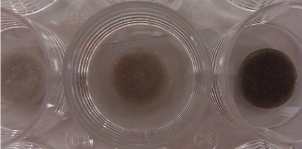
Researchers at Rice University and Texas Children’s Hospital (TX, USA) have developed paediatric heart-defect patches with carbon nanotubes that enhance electrical connections between cells. The patches are made of a sponge-like bioscaffold that contains microscopic pores and mimics the body’s extracellular matrix. They are infused with conductive single-walled carbon nanotubes that overcome a limitation of current patches in which pore walls hinder the transfer of electrical signals between cardiomyocytes. Cardiomyocytes are the heart muscle’s beating cells, which take up residence in the patch and eventually replace it with new muscle.
This invention could serve as a full-thickness patch to repair defects due to Tetralogy of Fallot, atrial and ventricular septal defects and other defects without the risk of inducing abnormal cardiac rhythms, according to the research team led by bioengineer Jeffrey Jacot and chemical engineer and chemist Matteo Pasquali. Results of the study have been published in the American Chemical Society journal ACS Nano.
Patch Degrades in Weeks or Months
The new heart-defect patch consists mainly of hydrogel and chitosan, a widely used material made from the shells of shrimp and other crustaceans. The patch is attached to a polymer backbone that can hold a stitch and keep it in place to cover a hole in the heart, the researchers explained. The pores make it easy for natural cells to invade the patch, which degrades as the cells form networks of their own. The patch, including the polymer backbone, also degrades in weeks or months as it is replaced by natural tissue.
Data show that once cells populate the patches, they cannot just synchronise with the rest of the beating heart because the scaffold mutes electrical signals that pass from cell to cell. That temporary loss of signal transduction results in arrhythmias, the researchers noted.
Nanotubes are able to fix the problem by enhancing the electrical coupling between cells that invade the patch, helping them to keep up with the heart’s steady beating. The insulating scaffold can delay the cell-to-cell signal further, but the nanotubes forge a path around the obstacles.
“We’ve been looking for a way to get better cell-to-cell communications and were concentrating on the speed of electrical conduction through the patch. We thought nanotubes could be easily integrated,” Jacot said, citing the relatively low concentration of nanotubes (i.e., 67 parts per million in the patches that tested best) as key. Previous attempts to use nanotubes in heart patches employed much higher quantities and different methods of dispersing them.
Jacot's team also found that a component they were already using in their patches – chitosan – keeps the nanotubes spread out. “Chitosan is amphiphilic, meaning it has hydrophobic and hydrophilic portions, so it can associate with nanotubes (which are hydrophobic) and keep them from clumping," Jacot said. "That’s what allows us to use much lower concentrations than others have tried.”
However, the toxicity of carbon nanotubes in biological applications remains a concern among experts. The fewer are used, the better, according to Pasquali. “We want to stay at the percolation threshold, and get to it with the fewest nanotubes possible,” he said. “We can do this if we control dispersion well and use high-quality nanotubes.”
The heart patches start as a liquid. When nanotubes are added, the mixture is shaken through sonication to disperse the tubes, which would otherwise clump, due to van der Waals attraction. Clumping may have been an issue for experiments that used higher nanotube concentrations, Pasquali added.
Fingernail-Sized Discs
The material is spun in a centrifuge to eliminate stray clumps and formed into thin, fingernail-sized discs with a biodegradable polycaprolactone backbone that allows the patch to be sutured into place, the researchers explained. Freeze-drying sets the size of the discs’ pores, which are large enough for natural heart cells to infiltrate and for nutrients and waste to pass through.
In addition, nanotubes make the patches stronger and lower their tendency to swell while providing a handle to precisely tune their rate of degradation, giving hearts enough time to replace them with natural tissue, said Jacot, who has a joint appointment at Rice University and Texas Children’s Hospital.
"If there’s a hole in the heart, a patch has to take the full mechanical stress,” Jacot said. “It can’t degrade too fast, but it also can’t degrade too slow, because it would end up becoming scar tissue. We want to avoid that.”
Source: Rice University
Image Credit: Rice University
Latest Articles
Arrhythmia, heart-defect patch, carbon nanotube, cardiomyocytes
Researchers at Rice University and Texas Children’s Hospital (TX, USA) have developed paediatric heart-defect patches with carbon nanotubes that enhance...


