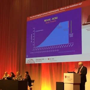
When it comes to imaging for ischaemic and non-ischaemic cardiomyopathy, clinicians should use their clinical acumen in order to decide which modality will lead to early diagnosis and treatment, said Prof. Petros Nihoyannopoulos, who spoke to HealthManagement.org after co-chairing a popular session on the topic at the European Society of Cardiology congress in Munich this week.
Patients with ischaemic or nonischaemic cardiomyopathy are common. Such patients may come to GPs and hospitals with symptoms, but symptoms are not very specific. Symptoms can be open and variable and include chest pains or breathlessness.
Dr. Nihoyannopoulos, who is Professor of Cardiology at Imperial College London, UK, emphasised that guidelines are only guidelines and not dogma. A good clinician needs to tease out what modality to use to lead to the diagnosis and treatment of the patient early enough. The choice of imaging modality needs to be guided by clinical suspicion and clinical presentation, such as if there is another systemic disease or skin or neurological lesions. All need to be put into perspective so that the imaging diagnosis can be refined early and not wait for late presentation
A key issue in cardiomyopathy is the presence of myocardial tissue viability, as the presence of viability will lead to treatment or re-vascularisation. In current guidelines this was attempted to be answered by the Surgical Treatment for Ischemic Heart Failure (STICH) trial, the only large-scale RCT on the subject, which unfortunately did not give an answer. Many imaging modalities that used viability tests were not included in this trial, such as MRI or PET, which does not reflect current clinical practice.
The most common modality to assess viability is stress echo and MRI with detection of fibrosis. Guidelines on these modalities will be implemented in the next updates, said Nihoyannopoulos . Every cardiomyopathy whether arrhythmogenic or idiopathic, has a typical phenotype, but each disease has a spectrum, so you may have a very mild form of infiltrative cardiomyopathy, amyloidosis or sarcoidosis or a very typical textbook type of phenotype that can be detected by any imaging modality. These are the easy cases, and there is little doubt that after echocardiography cardiac MRI is essential in order to make the diagnosis, noted Nihoyannopoulos. Coronary angiography is vital to detect anatomic coronary artery disease as opposed to just function. Again, this will be included in the updated guidelines, he added.
Source: HealthManagement.org
Image Credit: HealthManagement.org
Latest Articles
#ESC2018
When it comes to imaging for ischaemic and non-ischaemic cardiomyopathy, clinicians should use their clinical acumen in order to decide which modality wi...


