


Deep learning-based image reconstruction (DLIR) has emerged as a powerful tool in computed tomography (CT) to reduce image noise, decrease artefacts, and improve image quality, thus lowering the required radiation dose. DLIR algorithms are trained on low-dose CT data to produce high-quality images comparable to those obtained with routine or high-dose data. This approach has enhanced image quality in various CT applications, including cardiovascular, thoracic, and abdominal imaging, which rely on qualitative visual analysis.
However, the impact of DLIR on quantitative CT applications, such as coronary artery calcium scoring (CACS), remains under investigation. CACS, important for assessing ischemic heart disease risk, typically uses the Agatston method based on filtered back projection (FBP) images, which are noisy and limit dose reduction. While reducing noise, iterative reconstruction (IR) techniques tend to underestimate Agatston scores by 6–50%, limiting their acceptance for CACS.
Initial studies suggest that DLIR results in less underestimation of CACS (4–7%) than IR. Since DLIR offers varying levels of noise reduction, further evaluation is needed to determine the optimal settings for accurate CACS. A recent study published in Academic Radiology aims to systematically compare the impact of different reconstruction techniques (FBP, ASiR-V, and DLIR) on quantitative CACS in patients and assess DLIR's potential for dose reduction in a controlled phantom study.
Study Design and Methods
This retrospective, single-centre study, approved by the Institutional Review Board, included 100 adult patients who underwent calcium-scoring CT between June 2022 and May 2023, excluding those with coronary stents. An anthropomorphic thorax phantom with hydroxyapatite calcifications was used to simulate different chest sizes. CT scans were performed using a 256-detector-row CT with prospective ECG-triggering, 120 kV, and automated tube current modulation. Images were reconstructed with FBP, ASiR-V (30%, 60%, 90%), and DLIR (low, medium, high) techniques. Coronary artery calcium scoring utilised SmartScore 4.0 software. Image noise was measured using regions of interest in the phantom. Statistical analyses, including Bland-Altman plots and Friedman tests, were conducted with GraphPad Prism 9.0.0, using FBP as the reference. Significance was set at P ≤ 0.05, and agreement in CAC classification across reconstruction techniques was evaluated.
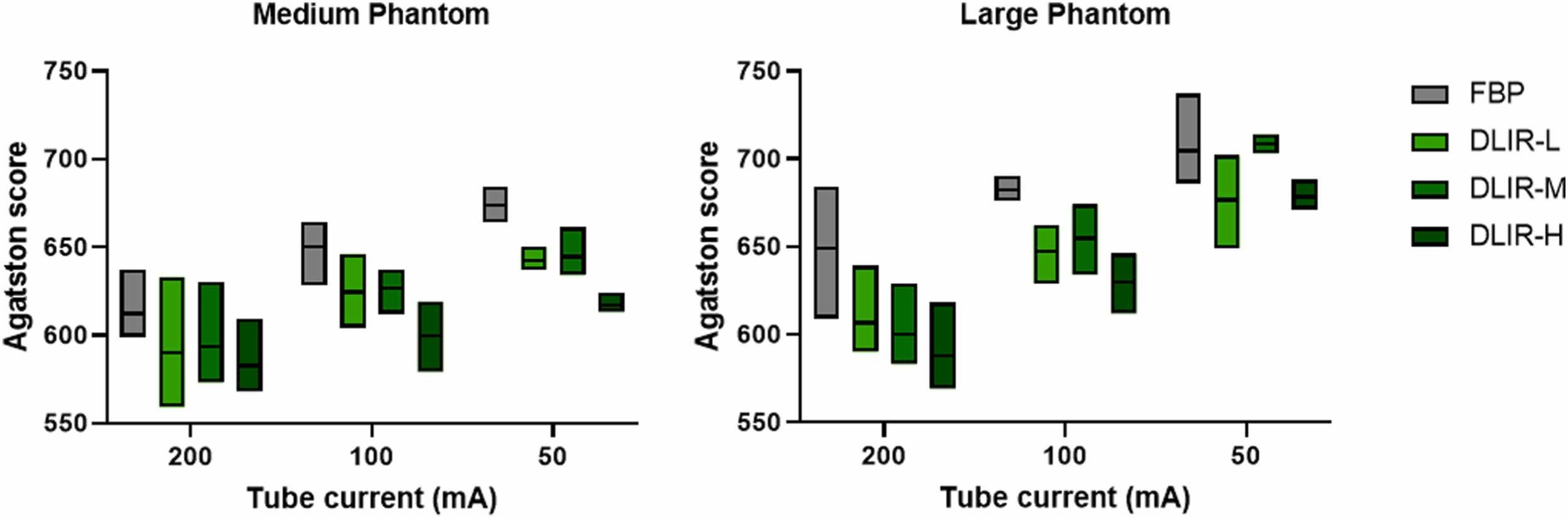
Image Credit: Academic Radiology
Patient Characteristics
The study included 100 patients with a mean age of 62 (±10) and 40% female. The average BMI was 26.5 ± 4.0 kg/m². Using FBP images as the reference, the median Agatston score was 31 (range 0–4744), with 30 patients having a score of zero. Calcium scores were minimal (1–10) in 11 patients, mild (11–100) in 20 patients, moderate (101-400) in 28 patients, and severe (>400) in 11 patients.
Impact of ASiR-V and DLIR on Quantitative CACS Results
DLIR showed better agreement with FBP for calcium scoring compared to ASiR-V, with the lowest mean bias in DLIR-L (-3.5) and the highest in ASiR-V 90% (-14.2). ASiR-V at 30%, 60%, and 90% strengths caused re-classifications of patients from non-zero to zero scores in six to seven cases, while DLIR at low, medium, and high strengths caused four, six, and eight such re-classifications, mostly from minimal to zero or mild to minimal scores. DLIR-L and DLIR-M did not reclassify moderate or severe risk groups, whereas DLIR-H reclassified one moderate case to mild.
Impact of DLIR on CACS and Radiation Dose Reduction
This study investigated the effect of deep learning-based image reconstruction (DLIR) on coronary artery calcium scoring (CACS) and its potential for radiation dose reduction in a retrospective patient cohort and a prospective phantom study. CACS is a critical marker for assessing cardiovascular risk and guiding preventive treatments like statins. While traditional filtered back projection (FBP) is the basis for understanding CACS's prognostic value, iterative reconstruction (IR) has shown underestimation issues. DLIR has emerged as a promising alternative, showing better agreement with FBP and less underestimation of CACS. The study uniquely combines a large patient cohort with a phantom study to analyse the influence of body size, dose levels, and reconstruction strengths on CACS results. We found that DLIR, especially at low and medium strengths, closely matched FBP, with minimal risk reclassifications.
The findings align with existing literature, demonstrating that DLIR-based CACS offers better accuracy compared to IR-based methods. DLIR reconstructions showed smaller biases and fewer discrepancies in risk classification than IR. The phantom study revealed that DLIR could effectively compensate for dose reductions, maintaining CACS results comparable to standard dose FBP reconstructions. This suggests that DLIR can enable substantial radiation dose reductions without compromising quantitative accuracy. However, the study's limitations include using DLIR and IR algorithms from a single manufacturer and a static phantom that does not account for cardiac motion or other clinical variations. Future prospective clinical studies are needed to confirm these results and further validate DLIR's efficacy in diverse patient populations.
Source: Academic Radiology
Image Credit: iStock