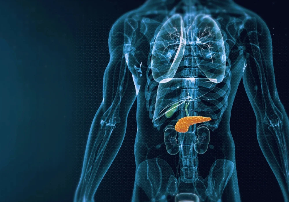
Magnetic resonance imaging (MRI) is increasingly used alongside computed tomography in pancreatic assessment due to superior contrast resolution and the absence of radiation. Technical advances have enabled high-resolution three-dimensional T1-weighted gradient echo (3D T1w GRE) acquisitions that improve anatomical detail but may reduce signal-to-noise ratio (SNR) and introduce artefacts when heavily accelerated. An evaluation at 3.0 T compared a deep-learning reconstruction (DLR) of the arterial-phase high-resolution 3D T1w GRE sequence with the standard reconstruction used in clinical care. Outcomes included overall image quality, artefacts, lesion conspicuity and quantitative measures such as SNR and lesion contrast-to-noise ratio (CNR). The findings inform how reconstruction choices can influence clarity of pancreatic anatomy and visibility of lesions during routine contrast-enhanced MRI.
Imaging Workflow and Reconstruction Approach
A single-centre, retrospective cohort of 32 patients was analysed from examinations performed between December 2021 and June 2022. All patients underwent contrast-enhanced pancreatic MRI at 3.0 T using a whole-body system with dedicated phased-array coils. Antispasmodic medication was administered before imaging to reduce peristalsis. The protocol included diffusion-weighted imaging and T1/T2 sequences before contrast, followed by gadolinium-based extracellular contrast injection at 0.1 mmol/kg with a saline flush. Arterial, portal venous and late phases were acquired in breath-holds. The arterial-phase sequence of interest used a 3D GRE with fat suppression, an acquisition voxel of 1.5 × 1.6 × 1.4 mm and a reconstructed voxel of 0.4 × 0.4 × 0.7 mm. Mean breath-hold for the arterial acquisition was 22 s.
Must Read: Improving Early Pancreatic Cancer Detection with DL
A vendor-supplied prototype 3D algorithm was applied offline to the same arterial-phase raw data to generate DLR images. The convolutional neural network estimates image noise and applies user-selected denoising; the denoising level was set at 75% based on prior experience and initial testing. Training used more than 10,000 image pairs with extensive augmentation to improve robustness for 3D reconstructions. Two radiologists, blinded to reconstruction type and clinical information, independently reviewed images on diagnostic workstations. They rated overall image quality and artefacts on 4-point scales, scored lesion conspicuity on a 3-point scale when lesions were present and counted lesions by location. Quantitative analysis measured SNR in the head, body and tail, and lesion CNR using standardised regions of interest. Statistical testing included paired t-tests for SNR and CNR, Wilcoxon signed-rank tests for ordinal ratings, weighted Cohen’s kappa for interobserver agreement and McNemar tests for sensitivity and specificity.
Image Quality Gains and Observer Agreement
The cohort included 16 women and 16 men with a mean age of 62 years. Across 24 patients, 38 lesions were identified, 8 patients had no lesions. DLR produced higher overall image quality scores than standard reconstruction for both readers. Reader 1 rated DLR at 3.23 versus 2.32, and reader 2 at 3.45 versus 3.03, with p < 0.01 for both comparisons. Artefacts were reduced with DLR for one reader (3.23 vs 2.81, p < 0.01) and were similar for the other reader (3.29 vs 3.19, p = 0.39).
Lesion conspicuity improved with DLR for both readers. Reader 1 reported 2.39 with DLR versus 1.88 with standard reconstruction, and reader 2 reported 2.18 versus 1.81, with p < 0.01 in both cases. Interobserver agreement also shifted favourably. For overall image quality, agreement rose from slight with standard reconstruction (κ = 0.09) to moderate with DLR (κ = 0.54). For motion artefacts, agreement improved from moderate (κ = 0.48) to substantial (κ = 0.69) with DLR. Agreement for lesion conspicuity was moderate with both methods (κ = 0.41 for standard reconstruction and κ = 0.29 for DLR).
Quantitatively, DLR increased SNR across all pancreatic subparts. In the head and neck, SNR rose from 147.93 ± 54.56 to 226.39 ± 88.61, in the body from 139.19 ± 51.27 to 214.59 ± 82.13 and in the tail from 129.07 ± 56.22 to 199.32 ± 91.02, with p < 0.01 for all. Lesion CNR improved from 11.15 ± 10.39 with standard reconstruction to 12.60 ± 12.38 with DLR, also with p < 0.01. These gains align with the algorithm’s design to reduce noise while preserving detail at near-isotropic resolution.
Lesion Detection and Practical Constraints
Reader-level sensitivity to detect at least one lesion per examination increased with DLR but without statistical significance in this cohort. Reader 1’s sensitivity was 1.00 with DLR versus 0.88 with standard reconstruction, and reader 2’s was 0.88 versus 0.83; both comparisons had p = 0.62. Specificity for reader 1 was lower with DLR than with standard reconstruction (0.43 vs 0.57, p = 0.48), while reader 2’s specificity was unchanged (0.86 vs 0.86). The evaluation therefore demonstrated clearer depiction and higher conspicuity without a measurable improvement in diagnostic performance at the examination level within the available sample.
Several factors frame interpretation. The work was single centre and retrospective, conducted exclusively on 3.0 T scanners, which may limit broader applicability given the prevalence of 1.5 T systems. The cohort was small with a limited number of lesions, constraining statistical power for diagnostic endpoints. The arterial-phase acquisition had a mean breath-hold of 22 s, which may challenge some patients. Case selection reflected practical considerations, including exclusion for incomplete coverage, breath-hold failure and unavailable raw data for reconstruction, which may introduce bias. Despite these constraints, the protocol maintained the same acquisition while altering only reconstruction, isolating the impact of DLR on image characteristics.
Applying deep-learning reconstruction to near-isotropic arterial-phase 3D T1w GRE pancreatic MRI at 3.0 T increased SNR and lesion CNR, reduced artefacts for one reader, improved overall image quality and enhanced lesion conspicuity. Interobserver agreement for image quality and artefact assessment also improved. Sensitivity gains were observed but were not statistically significant in this cohort, and specificity findings were mixed. For imaging teams, these results indicate that reconstruction choice can meaningfully influence visual clarity and confidence when reviewing contrast-enhanced pancreatic MRI without changing acquisition parameters. In settings where near-isotropic depiction and multiplanar reformats are important for evaluating pancreatic lesions and their relations, DLR offers a practical route to higher quality images that may support consistent assessment across readers.
Source: Insights into Imaging
Image Credit: iStock


