Artificial intelligence is reshaping breast imaging by addressing two persistent challenges: heavy reporting workload and variable diagnostic accuracy. Systems now show value in lesion detection, specificity and reading efficiency, with additional potential in individualised risk prediction, molecular subtyping and response assessment for neoadjuvant therapy. Evidence is strongest in population mammographic screening, where accuracy and workload gains have been broadly demonstrated. Applications in digital breast tomosynthesis, ultrasound and MRI are advancing but remain earlier in adoption or largely confined to research. Governance, cost-effectiveness and long-term clinical impact require further evaluation, and in current practice available tools are positioned as optional aids that radiologists can overrule. Careful implementation and ongoing outcome surveillance are therefore central to safe use.
Screening Mammography: Gains with Caveats
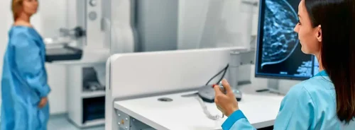
In digital mammography, multiple reader studies and prospective evaluations report improved accuracy and efficiency when AI supports reading. Measures such as AUC, sensitivity and specificity have risen without adding reading time, and in some comparisons AI as a standalone reader matched or exceeded many individual radiologists. Meta-analytic evidence across digital mammography and tomosynthesis indicates higher sensitivity but slightly lower specificity than human readers, with performance varying by study design and cohort.
Must Read: Improving Mammography Report Clarity with AI
Workflow impact is a central benefit. When used as a second reader, AI can reduce workload while maintaining non-inferior accuracy. Decision-referral approaches, in which AI triages normal and suspicious cases for human review, have shown improved performance over unaided reading and meaningful reductions in reading volume across large screening cohorts. Subgroup analyses describe better detection of invasive lesions, small tumours and findings in dense breast tissue. Early programme experience shows feasibility: AI-flagged cases not recalled by standard double reading have yielded additional cancers with only modest increases in recall.
Evidence from a large randomised screening trial demonstrated non-inferior cancer detection with substantial workload reduction when AI was used to triage cases. Hybrid strategies combining AI with targeted human review detected more cancers at similar workload levels, whereas fully autonomous reading did not outperform traditional double reading. Long-term outcomes, including interval cancer rates and downstream utilisation, were not available at the time of reporting.
Screening also benefits from risk assessment. A mammogram-derived deep learning model identified more high-risk patients than traditional tools among women undergoing supplemental MRI. Radiomic analysis of mammographic parenchyma associated with higher invasive cancer risk, including in false-negative and interval cancers and across black and white women, independent of density. These results support more accurate individual risk stratification that could inform personalised screening strategies. On balance, implementation appears advantageous and could partly replace one reader in double reading models, but programmes should pair deployment with post-market surveillance tracking recall, biopsy and detection metrics.
Beyond Mammography: DBT and Ultrasound in Practice
Digital breast tomosynthesis improves diagnostic performance but increases reading time and interpretative complexity. Contemporary systems can process DBT across vendors and have demonstrated notable reading time reductions while maintaining accuracy, supported by functions that highlight suspicious regions and generate synthetic views. Triage methods have reduced workload and improved sensitivity, and AI performance has approached human double reading in some settings, with particular utility in dense breasts. However, directly applying mammography-trained algorithms to synthetic mammograms may lower sensitivity, indicating the need for modality-specific tuning. Current evidence does not yet justify strong recommendations for DBT-specific AI as a standalone solution, though concurrent use during interpretation can shorten reading and improve accuracy.
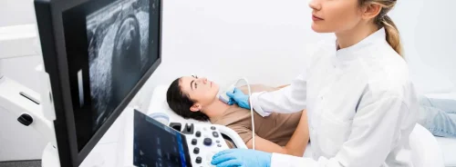
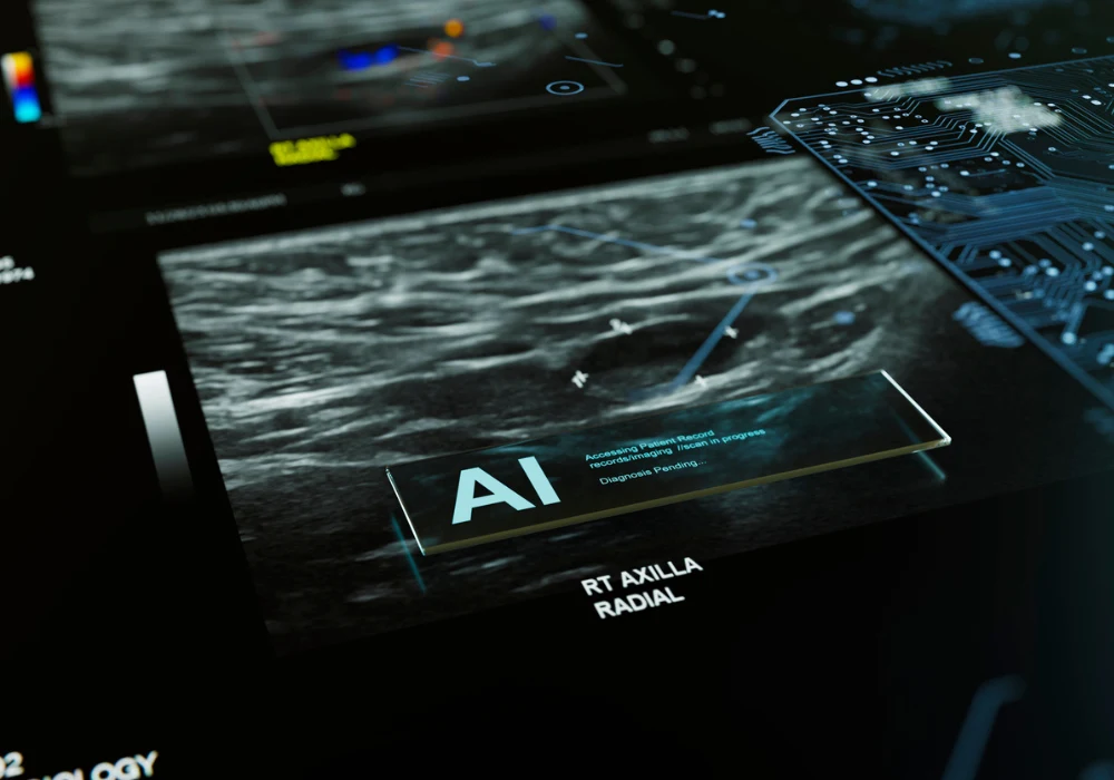
In ultrasound, AI targets lower specificity in older women, variability in acquisition and interpretation and time burdens. Computerised assessment of BI-RADS features has delivered high discrimination with fewer unnecessary biopsies. Deep learning, especially convolutional architectures, supports faster and more accurate malignant lesion classification, with performance comparable to radiologists and more consistent gains for less experienced readers. Embedding support within ultrasound devices can stabilise BI-RADS categorisation and reduce inter-reader variability, promoting standardisation across services.
Advanced ultrasound techniques such as shear wave elastography also benefit. Radiomics and machine learning models that combine B-mode and elastography outperform B-mode alone, and end-to-end deep learning that extracts elastography features automatically has shown high AUCs. AI has aided axillary staging by integrating elastography and B-mode features to detect metastatic nodes with strong discrimination and accuracy. Automated breast ultrasound radiomics shows promise in diagnosis, treatment evaluation and prediction of molecular markers, with further progress linked to multimodal data fusion, privacy safeguards and improved interpretability. Regulatory-cleared tools exist for lesion classification and biopsy reduction, but real-world post-implementation monitoring remains limited and should be prioritised.
MRI, CEM and LLMs: Promise with Limited Clinical Use
Most MRI applications remain research-oriented. Commercial CAD adds limited value for experienced readers, whereas newer AI models focus on triage and workload management. Retrospective analyses indicate that AI can detect a proportion of previously missed cancers and dismiss a meaningful share of normal cases while maintaining high sensitivity, with subsequent validations suggesting reduced biopsy rates. Workload reductions have also been reported with ultrafast sequences at high sensitivity, though performance can drop in screening-like datasets, highlighting gaps between development cohorts and real-world practice.
Risk and recurrence prediction on MRI is a second active stream. AI has classified Oncotype DX categories with high AUC and outperformed subjective assessments using background parenchymal enhancement to predict cancer onset. Models estimating five-year risk from a single maximum intensity projection in high-risk women have surpassed traditional tools. Radiomics-based virtual biopsy on MRI and contrast-enhanced mammography has achieved subtype discrimination with variable sensitivity and specificity by phenotype. For therapy planning, AI has predicted response to neoadjuvant treatment with promising AUCs that improve when clinical variables and longitudinal multimodal data are included. Despite these signals, clinical use beyond basic colour coding and registration cannot be recommended at present.
Contrast-enhanced mammography faces practical constraints related to radiation exposure, contrast reactions and lower specificity than MRI. AI development is at an early stage, with radiomics indicating potential for molecular subtype classification and exploration work showing synthetic contrast-enhanced images from mammography that resemble real images on qualitative review. Further architectural refinement, external validation and investigations into contrast dose reduction are needed before clinical testing. Large language models could support decision-making, report standardisation and guideline integration, and multimodal extensions may aid tumour classification and workflow, but domain-specific training and validation are lacking, so clinical use should be discouraged for now.
AI is advancing breast imaging by improving screening accuracy and efficiency and by opening pathways to personalised risk assessment and response prediction. Translation beyond screening is uneven, with DBT and ultrasound demonstrating practical benefits that need real-world validation and MRI and contrast-enhanced mammography largely anchored in research. Implementation should include robust governance and post-market surveillance focused on recall, biopsy and detection metrics. In clinical use, tools function as optional aids within radiologist-led decision-making, ensuring that algorithms support rather than supplant expert judgement.
Source: European Radiology
Image Credit: iStock


