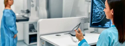
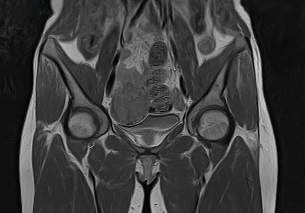
The advent of quantitative MRI techniques marks a transition in the clinical management of uterine cancers, notably endometrial cancer (EC) and cervical cancer (CC). These malignancies, though originating in the same anatomical region, differ significantly in epidemiology, pathology and clinical behaviour. Historically reliant on morphological imaging and histopathological data, oncological diagnostics now benefit from advanced imaging biomarkers that offer functional and molecular insights. Quantitative MRI, incorporating modalities such as Dynamic Contrast-Enhanced MRI (DCE-MRI), Diffusion-Weighted Imaging (DWI) and Magnetic Resonance Spectroscopy (MRS), bridges the gap between non-invasive imaging and personalised treatment strategies.
Enhanced Diagnostic and Prognostic Insights with DCE-MRI
DCE-MRI allows clinicians to quantify physiological parameters of tissue microcirculation, a process central to tumour biology. Parameters such as Ktrans, Ve and Vs offer key insights into perfusion and vascular permeability, providing vital clues about tumour aggressiveness. These biomarkers have proven superior to conventional T2-weighted imaging for detecting critical features like myometrial or cervical invasion in EC and CC.
Must Read: Ultrasound vs MRI in Cervical Cancer Staging
In EC, DCE-MRI supports risk stratification by revealing perfusion patterns linked to histological and molecular subtypes. For instance, lower Ktrans and Ve values correlate with poorly differentiated tumours, while higher values are associated with better-differentiated, low-grade cancers. Such quantitative assessments aid in distinguishing high-risk subtypes and guiding surgical or adjuvant therapy decisions. DCE-MRI has also shown promise in identifying TP53 mutations and predicting treatment resistance, which is critical for tailoring therapeutic strategies.
In CC, where traditional biomarkers are limited, DCE-MRI contributes significantly to predicting treatment outcomes. Poor perfusion and hypoxia—detectable through low enhancement values—are often indicators of radiotherapy resistance. Thus, early identification of low-perfusion tumours could prompt treatment modifications to improve prognosis.
Diffusion-Weighted Imaging and Intravoxel Incoherent Motion
DWI, particularly with high b-value sequences and the Intravoxel Incoherent Motion (IVIM) model, is instrumental in differentiating between benign and malignant lesions and assessing therapeutic response. Apparent diffusion coefficient (ADC) values, derived from DWI, tend to be lower in aggressive or poorly differentiated tumours due to increased cellularity that restricts water movement.
The IVIM approach enhances diagnostic specificity by distinguishing tissue diffusion from microvascular perfusion. Parameters such as true diffusion (D), pseudo-diffusion (D*) and perfusion fraction (fp) are particularly valuable. In EC, lower ADC and D values have been associated with higher tumour grade and stage. Similarly, in CC, these parameters help predict lymph node involvement and assess response to neoadjuvant chemotherapy.
Importantly, IVIM parameters show strong correlations with histological markers such as tumour-infiltrating lymphocytes and lymphovascular invasion. This alignment between imaging and pathology supports their use in refining clinical staging and therapeutic decision-making, although further standardisation in acquisition protocols and parameter fitting is necessary to enhance reproducibility.
Magnetic Resonance Spectroscopy: A Metabolic Window into Tumour Biology
MRS offers a unique perspective by non-invasively quantifying tumour metabolites. Signals from choline-containing compounds and lipids, prominent in uterine malignancies, serve as potential biomarkers for diagnosis, grading and treatment response. Elevated choline reflects high cellular turnover, typical of malignant tissues, while lipid accumulation is increasingly associated with poor prognosis and therapy resistance.
In EC, high choline signals correlate with features such as increased tumour volume, high Ki67 expression and lymph node metastasis. Lipid peaks, especially those resonating at 1.3 ppm, have been proposed as indicators of tumour hypoxia and aggressiveness, particularly in CC. MRS thus not only aids in preoperative risk stratification but also offers potential for monitoring metabolic shifts in response to treatment.
Despite its potential, MRS faces barriers to widespread clinical use. Its implementation demands high-field MRI systems (≥3T), expertise in spectral acquisition and rigorous standardisation. Nonetheless, as motion artefact mitigation techniques improve, MRS is becoming more accessible for pelvic imaging, expanding its diagnostic and prognostic utility in gynaecological oncology.
Quantitative MRI represents a transformative advancement in the personalised management of uterine cancers. Techniques like DCE-MRI, DWI and MRS transcend conventional anatomical imaging by providing functional, structural and metabolic biomarkers that enrich diagnosis, prognostication and treatment planning. By integrating these modalities into clinical workflows, oncologists can achieve a more nuanced understanding of tumour biology, enabling tailored therapeutic strategies that align with individual patient profiles. With due standardisation and validation, quantitative MRI is set to become a central pillar of precision medicine in uterine oncology.
Source: Insights into Imaging
Image Credit: iStock


