HealthManagement, Volume 6 - Issue 5, 2006
Practical Guidelines for a Successful 3D Imaging Service
Authors
Gordon J. Harris
Director, 3D Imaging
Service
Massachusetts General
Hospital
Associate Professor of
Radiology
Harvard Medical School
Boston, MA
USA
Matthew A. Barish
Director, 3D and Image
Processing Center
Brigahm and Women’s
Hospital
Assistant Professor of
Radiology
Harvard Medical School
Boston, MA
USA
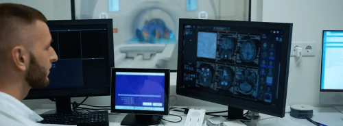
PACS soft-copy reading, as with film reading, has traditionally been limited to viewing the transmission of cross-sectional images in fixed orientations as provided by the original scan. Improvements in the speed, usability and affordability of 3D workstation hardware and software, and improvements in integration of 3D software within PACS, means it’s now possible to apply computer image processing techniques for 3D imaging and CAD within routine clinical workflow to provide 3D imaging within a PACS environment.
MDCT has opened new possibilities and increased demand for 3D imaging. Rapid acquisition using thin slices and improved rendering algorithms facilitates exquisite 3D images and reformats. However, review of the ensuing large numbers of images from modern MDCT and MR scanners, often hundreds or even thousands of slices per study, pose significant challenges to radiologists’ efficiency. Having 3D capability available for diagnosis and surgical planning allows radiologists and referring physicians to get a summary view of the entire anatomy in a few concise, anatomically clear 3D views, and then refer back to the original 2D data for comparison and confirmation. Thus, 3D reconstruction is more frequently becoming a valuable technique to summarise in a concise and clear way the overwhelming number of slices produced by modern CT and MR scanners.
3D medical imaging has revolutionised both radiological diagnosis and surgical planning. 3D reconstructions provide life-like views that can quickly summarise the relationship among anatomic structures for planning surgical procedures before and within the OR. Benefits include decreased exploratory time in the OR, less damage to healthy tissues, and a lower risk of complications for the patient, all of which contribute to reduced surgical morbidity.
How Can 3D Reduce Costs?
Increased diagnostic sensitivity across all specialties and the likelihood of shorter OR time per procedure contribute to reduced costs. Thus quality of care is greatly enhanced, often with a decrease in cost compared to alternative procedures. For example, prior to the establishment of 3D imaging services at our institutions, standard protocol for assessing laproscopic living renal donor candidates involved a catheter angiogram to assess the renal vasculature (number, location, and relation among renal arteries and veins) plus an intravenous pyelogram (IVP) to assess the configuration of the ureters and renal collecting system.
These procedures involved exposing the patient to significant health risk involving anaesthesia, catheterisation, as well as high radiation dose, and several thousand dollars in costs for both procedures. Through 3D imaging post-processing of high-resolution CT scans, the current protocol involves a CT angiography scan (CTA) to assess the renal vasculature in place of the catheter angiogram, plus a delayed phase CT urogram (CTU) which replaced the IVP, and both the CTA and CTU are acquired in a single outpatient CT contrast study (see Fig. 1).
Thus, a single, contrast-enhanced CTA/CTU outpatient procedure, costing a few hundred dollars with minimal risk and exposure to the patient, has replaced two expensive and invasive procedures costing several thousand dollars and exposing an otherwise healthy patient to significant risk. Ultimately, 3D imaging provides patients with improved care, significantly lower risk and cost and improved diagnostic clarity.
Potential Uses for 3D Imaging
3D imaging services have become an integral part of the clinical workflow for radiologists and referring clinicians in many hospitals as they can process exams for a variety of sub-specialties including vascular, orthopaedic, chest, breast MR, GI/GU, emergency and paediatric exams. In addition, a variety of neurology, neurosurgery and oral-maxillofacial exams are reconstructed with3D imaging, including brain surface renderings for neurosurgical planning, CTA for stroke and aneurysm imaging, brain tumour volumetric measurements, imaging of craniosynostoses and complex facial fractures/trauma.
3D reconstructions are also essential to vascular imaging. For example, aortic aneurysm, liver resection and living liver and renal donor assessments are greatly aided by pre-surgical 3D imaging of CT or MRA vascular evaluation and treatment planning on computer rather than exploration in the OR. 3D imaging is also effective in assessing pancreatic cancer and associated vascular involvement and for virtual endoscopy, such as colonography or bronchography.
Many complex 3D exams, CTA for example, require 45 to 60 minutes or more to process all the associated views, while other less complicated 3D exams may take only 15 to 30 minutes each to process. If the clinical volume at a facility is sufficient, it is cost-effective to have one or more dedicated 3D technologists process these exams or alternatively outsource the 3D image processing.
What to Look for in a 3D Technologist
A dedicated 3D technologist should be hired as an experienced, certified CT or MR technologist who can work closely with the radiologists and referring physicians to create 3D protocols that generate the most useful views for each type of exam. The 3D technologist must understand cross-sectional anatomy and pathology in-depth, as well as scan artifacts, and must learn to operate complex computer software. The training period for a dedicated 3D technologist thus ranges from 3 to 6 months at our institutions.
One consideration in determining the cost-effectiveness of the service would be the cost of not having a dedicated 3D imaging service. For example, if a radiologist spending a few hours per day performing 3D processing would be diverted from reading additional clinical cases, which would have generated added revenue, and the facility might need to hire additional radiologists to read exams. Likewise, if CT or MR technicians were to perform 3D imaging between scans, it would slow down scanner throughput, decreasing revenue. A dedicated staff for 3D imaging services can provide an efficient and consistently processed set of 3D views. By having a dedicated 3D technologist processing exams, it is also possible to create greater uniformity and consistency across exams, as opposed to having 3D images produced by a variety of radiologists or technologists who are not trained to produce a clinically determined set of 3D imaging protocols.
Requirements for a Successful 3D Imaging
Service
Successful implementation of a 3D imaging service requires that clinical workflow, protocols, billing and staffing issues are established in a way that enables routine clinical 3D imaging with rapid turnaround and consistent, high-quality, clinically-relevant post-processed images. The 3D imaging services at our institutions work closely on an ongoing basis with hospital billing and compliance, coding and PACS to establish workflow, and with the radiologists, referring clinicians, CT and MR scan departments to establish clinical 3D and scan protocols optimised for clinically useful 3D imaging.
Conclusion
3D imaging in radiology has had a major clinical impact in the past decade in routine clinical practice. However, there is a cost and a learning curve involved in producing optimal clinical 3D imaging. The benefits of implementing 3D imaging services can include improved confidence in diagnosis and treatment/surgical planning, reduced cost and invasiveness of procedures, pre-surgical planning rather than exploratory surgery and ultimately less risk to the patient and improved patient care.


