


Accurate patient positioning is crucial in clinical imaging workflows, such as magnetic resonance imaging (MRI) and computed tomography (CT), to obtain high-quality images, ensure diagnostic accuracy, optimise radiation doses, and enhance patient comfort and safety. This accuracy is significantly dependent on the precise estimation of the patient's height and weight. Despite its importance, there are no widely established methods for these estimations, often leading to reliance on patient-provided or previously recorded data, which can be inaccurate. The advent of three-dimensional (3D) camera systems offers a promising solution for automatically and accurately measuring patient height and weight, thereby improving clinical imaging workflows.
Importance of Accurate Patient Positioning
Accurate patient positioning in imaging workflows is essential for several reasons. Firstly, it ensures high-quality images, which are crucial for accurate diagnoses. Mispositioning can lead to suboptimal images, potentially resulting in misdiagnosis or the need for repeat scans, exposing patients to additional radiation. Secondly, correct positioning enables the comparability of images over time, which is vital for monitoring disease progression or treatment efficacy. Additionally, optimised radiation doses are critical in CT imaging to minimise exposure while maintaining image quality. Lastly, accurate positioning enhances patient comfort and safety, particularly for those who are severely ill or immobile.
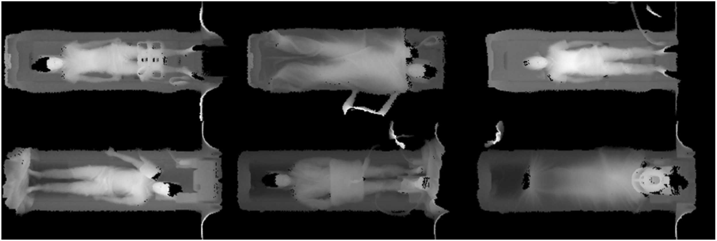
Image Credit: Academic Radiology
Accurate height and weight estimation play a pivotal role in patient positioning. These measurements are vital for calculating the Specific Absorption Rate (SAR) in MRI and the contrast dose in both MRI and CT. However, in clinical practice, height and weight are often not measured before scans, relying instead on estimates from the patient or medical technologists, which can be imprecise.
Challenges in Height and Weight Estimation
The primary challenge in height and weight estimation lies in the absence of standardised methods. Typically, measurements are not taken directly due to high patient throughput or the patient's condition, leading to reliance on inaccurate estimates. Methods proposed by Lorenz and Crandall involve additional measurements like mid-arm circumference, which are complex and not always feasible.
Medical technologist estimates have been shown to be less precise, highlighting the need for automated and accurate methods. The intense workload in radiology departments further necessitates automation to save time and optimise the scanning process, enabling more scans per day.
3D Camera Systems: A Promising Solution
3D camera systems offer a promising solution for accurate and automatic patient height and weight estimation. Initially implemented for patient auto-positioning in CT, these systems have recently been adapted for MRI. The myExam™ 3D Camera by Siemens Healthineers is an FDA-approved device integrated within the MR environment for clinical use. It uses infrared time-of-flight technology to acquire depth images and generate a 3D surface contour map, detecting anatomical landmarks for auto-positioning and estimating height and weight.
Recent studies have explored height and weight estimation using machine learning approaches with 3D camera images. A novel deep learning (DL) algorithm proposed by Tamersoy et al. addresses this estimation problem using a multi-stage training approach on a large dataset, ensuring accuracy and generalisation. This algorithm is now available in myExam™ 3D camera technology.
Evaluation of 3D Camera-Based DL Algorithm
A study was conducted to evaluate a DL-based 3D camera algorithm's precision in estimating patient height and weight during clinical routine examinations on a low field 0.55 T MRI. The study hypothesised that the 3D camera's estimates would demonstrate higher accuracy and agreement with actual measurements compared to medical technologist estimates.
The study involved 148 patients, with height and weight measured using a calibrated scale and visually estimated by medical technologists. The 3D camera captured depth images, processed by the DL algorithm, to estimate height and weight. The results showed that the 3D camera-based method significantly outperformed medical technologist estimates in both accuracy and precision for height and weight.
Integrating 3D camera systems with deep learning algorithms in clinical imaging workflows significantly advances patient care. These systems provide accurate height and weight estimations, essential for optimal patient positioning, SAR calculations, and contrast dosing in MRI and CT. The study demonstrated that the 3D camera method outperforms traditional estimation methods, offering improved diagnostic accuracy, patient safety, and workflow efficiency. As the technology evolves, it promises further enhancements in clinical imaging practices, ensuring better patient outcomes and streamlined radiology operations.
Source: Academic Radiology
Title Image: iStock


