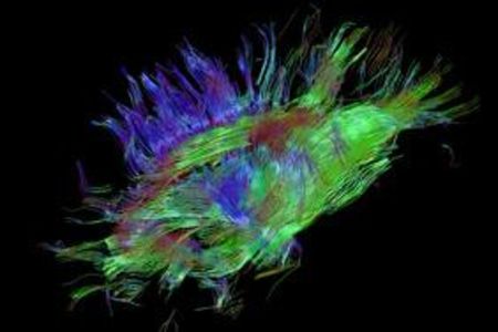
Researchers at the Eindhoven University of Technology have developed a new software tool, which converts MRI scans of the brain into three-dimensional coloured images of nerve structures.
The new technology gives physicians a complete picture of the winding roads and their contacts, enabling them to easily study the wiring of the brains of patients, without having to operate. The tool, which was developed by TU/e researcher Anna Vilanova, with her PhD students Vesna Prčkovska, Tim Peeters and Paulo Rodrigues, is based on a recently developed technology called HARDI (High Angular Resolution Diffusion Imaging). The MRI measuring technique for HARDI was already there but the research team took care of the processing, interpretation and interactive visualisation of these very complex data, leaving the physicians with a clear picture ready for analysis.
The tool accurately shows the location of the main nerve bundles in the brain, which is important for brain surgeons to know in advance, in order to avoid damaging them. The technique may also yield many new insights into neurological and psychiatric disorders.
Bart ter Haar Romenij, professor of Biomedical Image Analysis, at the Department of Biomedical Engineering, believes that the accuracy of the tool is a great step forward and expects that the new technique can be ready for use in hospitals within a few years, following validation and further improvements in speed of processing. The research conducted was supported by the NWO (Dutch Organization for Scientific Research).


