HealthManagement, Volume 15 - Issue 3, 2015
Portable Ultrasonic Scanning systems provide two-dimensional (2D) images of soft tissue and moving structures (eg, heart, fetus) for a variety of clinical applications. Depending on the transducer and calculation packages available, portable scanners can be used for abdominal, obstetric/gynaecologic, urologic, smallparts (eg, thyroid, prostate, breast), cardiac, and other examinations. Portable systems typically weigh less than 23 kg (50 lb), and are compact and lightweight enough to be carried by hand between exams.
Key Considerations
• Portable Ultrasound (US) systems come in three different configurations: (1) Handheld, (2) Tablet computer or (3) Laptop computer. Handheld US scanners are compact enough to be held in one hand during use while the transducer is held in the other hand. Tablet-style US systems have a similar user interface that includes an imaging display screen and a variety of controls activated through a touch screen. Laptopbased US systems typically have the most user-adjustable controls and imaging capabilities and are the most similar in function to full size, cart-based US systems.
• When purchasing any type of US system, facilities need to consider six basic issues: functions and features, cost, ease of use, upgradeability, image storage, and customer support.
• For portable US systems, additional considerations include size, weight, transducer options, and the availability of advanced imaging modes.
• Some portable systems may include additional transducers to facilitate more specialised cardiac, vascular, endovaginal, endorectal , or small - parts diagnostic procedures.
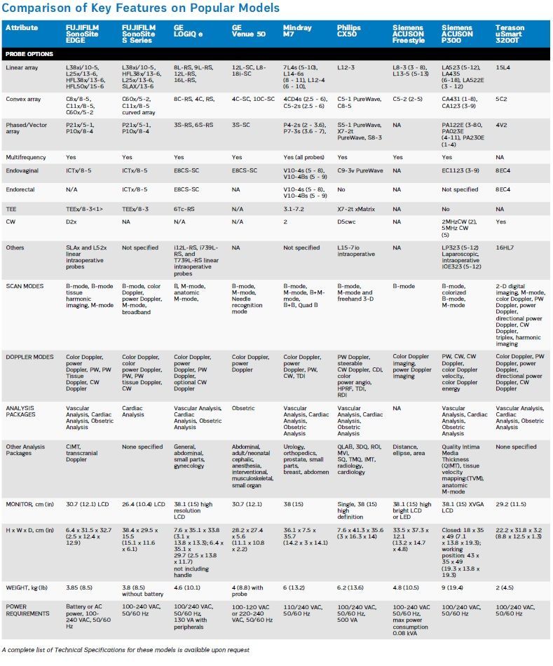
• A variety of transducers (also called “probes”) are typically available for use with portable systems. Many of the probes that are currently offered on popular systems are listed in the comparison table below.
• A number of portable US systems now offer advanced scanning features such as harmonics, Doppler colour flow mapping (CFM), and three-dimensional (3D) imaging. A few systems include additional advanced features that were typically only seen on full-size US systems, until recently.
Other Considerations
The availability of relatively inexpensive and easy-to-use portable ultrasound scanners has led to the technology b eingadopted by numerous non-imaging medical professionals for a wide range of point-of-care (POC) applications. These include anaesthesiology, endocrinology, rheumatology and sports medicine. Manufacturers have recognised that the POC ultrasound market is growing and they have begun to produce ultrasound systems that are designed to meet the demands of specific POC applications.
Many portable scanners now include Doppler capability to determine the direction and speed of blood flow. Doppler capabilities may include spectral Doppler, either continuous wave (CW) or pulsed wave (PW). Harmonic imaging is also available on some port able US scanners. Harmonic Imaging (HI) is a version of B-mode that, in many cases, improves image quality over that provided by conventional B-mode imaging.
Before purchasing a portable ultrasound scanner, buyers should consider scanning system functions and features relative to the number and types of procedures to be performed and choose probes and calculations packages accordingly.
Service and Support Information:
• Typical Warranty Coverage: 1 - 5 Years
• Estimated Service Life: 7 Years