HealthManagement, Volume 16 - Issue 4, 2016
INTERVIEW WITH GÁBOR FORRAI, EUSOBI PRESIDENT
What were some of the highlights of the European Society of Breast Imaging 2016 annual scientific meeting?
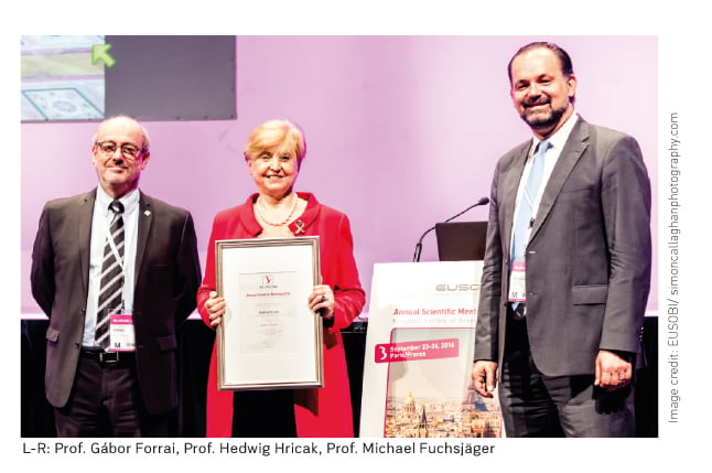
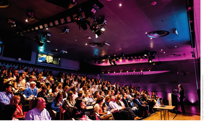
It was a fantastic meeting, with 730 attendees, the highest ever. Popular sessions included the BI -RADS workshop for standardising reporting in breast imaging and screening. The sessions on diffusion MRI were also very popular, as MRI is increasingly used to screen women with a high risk of breast cancer and also in diagnostics. There was an excellent session on managing B3 lesions (BI -RADS assessment category: Probably Benign). These borderline lesions are challenging to manage.
There was an interactive multidisciplinary team (MDT ) session, played in front of the audience as in a hospital, including discussion about clinical cases. As breast diagnostics and therapy is a multidisciplinary issue it’s impossible to decide on diagnostics and therapy alone. All these professions (radiologist, surgeon, oncologist) have to work together, therefore MDT meetings are obligatory in many countries. There is evidence that if there is a team decision patient management will be better, and in many cases after the team meeting there is a change of the therapy plan. A lot of the breast radiologist’s time is spent in team meetings, and this intellectual professional “thinking together” is an important addition to the diagnosis. Meetings are comparatively cheap —it’s not only the machines that can provide and add to the diagnosis and planning.
See Also: The Importance of Breast Screening: Zoom On Gabor Forrai
What’s current best
practice to manage borderline lesions?
There are some new features such as using MRI in order to try to predict the potential behaviour of these lesions. The question always is whether to operate or to observe. There is always a biopsy, which precedes this decision or issue, so if the biopsy confirms a lesion as B3 with malignant potential, the decision is always whether to excise something that may never cause harm to the woman, and the issue here is over-treatment or whether to schedule follow up. Here the risk is that the patient may not have regular follow up because she’s forgotten or moved and then we may face a breast cancer, so that means under treatment. We are always balancing between these two situations with B3 lesions. Breast radiologists can perform image-guided excisions of these lesions. I think that in a few years interventional therapy will be performed by breast radiologists for small breast cancers as well. It is already practised in some countries like Japan, eg for very old people who cannot undergo anaesthesia. It is not yet a routine method yet, as it is not allowed in every country.
Regarding information for women on mammographic screening, there is a lot of information and sometimes misinformation in the mass media and on the internet. How can this be improved?
This is mainly communication and public relations work, to reach the public with specific information to make them understand and convince them that the information is sound enough to be followed. Public relations work and doctors are fields apart, so there is a need for intermediates like the press and professional organisations to communicate. In the breast health field it is especially important, because there are fewer problems regarding communication regarding eg the liver and spleen because nobody thinks they know all about it, or gives hints about these lesions. In breast imaging misinformation is a huge problem. Because of this EUSOBI published recommendations for women’s information about mammography (Sardanelli and Helbich 2012), which are also intended to be read by referring clinicians. EUSOBI also published a paper with recommendations for women’s information on MRI (Mann et al. 2015).
This year the theme of the International Day of Radiology is breast imaging, EUSOBI , in collaboration with the U.S. Society of Breast Imaging, European Society of Radiology and Europa Donna, has published Screening & Beyond, which is free to download (internationaldayofradiology.com/publications). Aimed at women, the publication includes EUSOBI ’s recommendations for women’s information on mammography, MRI , ultrasound and image-guided breast biopsies. There are also chapters on standards and quality, research, the history of breast imaging, the role of the radiographer as well as information from Europa Donna. EUSOBI has strong links with 30 national breast imaging societies in its National Societies Network. We recommend that the national societies translate the women’s information materials into their own languages.

Should tomosynthesis be the new standard for mammographic screening programmes?
It is certainly an excellent imaging method. Already there are scientific articles that prove its superiority compared to simple digital mammography. However, the data are not enough to say that screening should be done with tomosynthesis. Many screening centres are doing an additional tomosynthesis 1 or 2 views to the standard views. Even standard views can be replaced by tomosynthesis with some special techniques. However, we have to have the data, the evidence, to prove it scientifically, and statistically. This slows down the initiation of a new and very good technique, but on the other hand avoids the risk that a technology may later prove not to be successful. I think tomosynthesis is good enough and will prove its superiority, but we need time and data.

How can rates of false positives and false negatives be improved?
There is always a balance. If the screening method is sensitive but not specific enough, it finds a lot of things, both positives and negatives, but will result in a lot of lesions. If the method is not sensitive enough, it means it will not find enough cancers. There is always a difficult balance between finding most of the cancers but not finding most of the benign lesions. There are no 100% perfect methods in the world, either for medical or non-medical issues. We have to realise that this phenomenon exists, that there will be always some positives and false negatives. The question is how to deal with it, and how the public will deal with it. Most important is that we use very quick and straightforward methods to prove that a lesion is false positive or true positive and the same for the negatives. We should not keep women waiting long weeks between screening, additional diagnostic tests and biopsy until the result is known, as it is damaging psychologically. With one-stop clinics, patients are recalled after screening, and have additional tests and, if needed, most of the biopsies on the spot, and receive the results within days.

As radiologists we need time and empathy to deal with false positives. We cannot avoid them, and we have to communicate with the woman what the probability is of the lesion being cancer. When you call back a patient, usually it’s by phone, it is much discussed with administrative or medical staff what to say to them. We usually say “We have found a lesion in your breast, which can be a simple asymmetry or something else." If the result is positive, we quietly inform the woman, that there is a lesion and that a therapy will be needed. You cannot avoid false positives, but communication and time are key.
Will computer-aided detection and diagnosis this play a bigger role in the future in breast imaging?
Definitely. Already artificial intelligence and data mining are taking more of a role as prices come down and products are more readily available. Such technology will help radiologists’ work, especially as there are many capacity problems in healthcare. But these technologies will not replace the decision maker. Computer-aided detection in mammography is almost 15-20 years old, and even if you use the latest technology, you get quite frequently some false positive marks made by the computer. Radiologists never make so many false positive marks, so the radiologist has to control the technology. Computer-aided detection is not adequate to replace the human radiologists, the scientific eye and experienced brain, but it assists.
EUSOBI’S education course on digital breast cancer screening has the objective that radiologists “ensure the correct balance between detection, specificity and the associated efforts and costs.” How do radiologists achieve this balance in practice?
We would like to decrease the personal "creative" approach towards diagnostics and to increase protocol-based standardisation. Of course there are cases when we have to depart from the protocol and do something additional or different. But first of all radiologists must know the protocol, and EUSOBI is working on protocols and flowcharts that show the steps to follow. In breast imaging there are so many lesions and this is such a dangerous disease that the only way forward is to propagate to work in a protocol-based way. If it is done this way, it will be less costly, more cost-effective and will decrease workload.
You are a forensic radiologist as well as a breast radiologist. What does this involve?
Forensic radiology is between medicine and law. I took on the role of a forensic expert and made the juridical exams in Hungary, as there are very few breast experts doing it. Such cases should not be judged by a physician who is not practising breast radiology, which has been the case in some malpractice issues. We need experts from the same field. There is a different logic in breast imaging and screening, as we are working from statistics, from probability, and there are many more lesions in the breast than in other parts of the body. In 80% of cases the question is an overlooked cancer: what would have happened if it had been diagnosed earlier? It’s an interesting mental and logical challenge.
This interview will appear in HealthManagement’s issue with a cover story on healthcare without borders. What does this concept mean to you as a breast radiologist?
It means that there is one proven method in the whole world for screening breast cancer in normal risk women, which is mammography, and that is what EUSOBI ’s 5 points against breast cancer emphasise (see box). Mammography is already without borders, as everywhere it is the same. Millions of women’s data from screening programmes have been collected in the world for 45 years, and the outcome of these data is used in other millions of women in the same and other parts of the world. Of course there are capacity and equipment accessibility variations between Africa and the USA , even between Europe and the USA that have to be taken into account by creating local protocols. Mammography is the most proven of all medical and diagnostic acts. It has the most scientific proof, even more so than a regular thoracic x-ray that is performed every day in hospitals
EUSOBI 5 Points
Against Breast Cancer
- In Europe, there are slightly different breast screening protocols
in every country, but one statement is uniform: mammography saves lives and
life quality – it can decrease breast cancer death by up to 40%.
- One in five breast cancers occur in women below 50, so
screening should start before this age, at least at 45.
- 45% of breast cancers and related deaths occur in women
at 65 and over. One in three breast cancers occur in women over 70, so
screening should not stop at this age, but continue at least up to 75.
- If a woman has breast symptoms, the breast radiologist is
the doctor to meet – he/she integrates all diagnostic techniques in order to
give the most accurate information on breast health status e. g. cancer
disease.
- The woman’s risk profile matters: personalized screening
protocols may include ultrasound and/or MRI – but should always be interpreted
together with mammography.


