HealthManagement, Volume 17 - Issue 5, 2017
Breast cancer screening and the breast imagers of the future
The future of breast examinations is changing. More and more screening methods are being invented as well as an influx of young radiologists hoping to make a difference. Members of EUSOBI tell us how.
Breast cancer is the most common female cancer and the 2nd leading cause of female cancer deaths worldwide with its incidence increasing. According to the Office for National Statistics and the Public Health England in 2016, 100% of patients diagnosed with breast cancer at stage I survived the disease for at least one year compared to only 63% of patients diagnosed at stage IV. Furthermore, breast cancer mortality is highest among women who are not screened regularly and consequently present with advanced cancers. This highlights the necessity for early diagnosis to improve survival outcomes of breast cancer patients.
The overarching goal is to detect breast cancer as early as possible. To date there are several breast imaging tools available including digital and contrast-enhanced mammography and digital breast tomosynthesis (DBT ), breast ultrasound (US) and magnetic resonance imaging (MRI ). Each of these techniques has certain advantages but also limitations.
Numerous randomised controlled trials have shown that screening with mammography enables early breast cancer detection, reduces breast cancer related mortality and thus has been implemented in many healthcare systems over the past three decades. Although mammography is the mainstay for screening, its sensitivity is limited and it is particularly less accurate in a sub-group of women with dense breasts. Supplemental imaging with other modalities such as DBT , US and MRI may lead to the detection of breast cancers that are not visible on mammography. The combined use of mammography (or DBT ), US and MRI could be the most sensitive approach to detect all breast cancer early, but at the same time this approach increases unnecessary recalls of women without cancer and comes at very high healthcare costs. Therefore, in the near future it is expected that mammography alone will not remain the primary screening protocol for all women; other modalities will gain a place.
Currently, breast MRI is considered the most accurate imaging method, detecting cancers that are not (yet) visible on other available imaging modalities. Moreover, breast MRI primarily detects biologically more aggressive breast cancers earlier whereas mammography is more biased towards detecting more indolent cancers. MRI is however relatively expensive, has limited availability and it is not tolerated by all women.
Because breast cancer continues to be a major cause of cancer related deaths in women, the search for improved breast cancer screening methods continues. Per definition, a screening tool should be effective, feasible and affordable because resources are limited in terms of cost and availability of breast radiologists.
In order to balance the costs and benefits of screening, screening research is turning towards personalisation of screening practice with different modalities offered with respect to an individual woman’s risk factors for developing breast cancer, such as family history, breast density and medical history. Moreover, in the era of precision medicine, screening tests should aim at identifying several hallmark capabilities that cancers acquire during their development [1, 2], in order to reduce the risk of overdiagnosis and overtreatment. In this context, MRI has proven to be a versatile and precise imaging technique that can simultaneously assess a multitude of functional cancer-related processes. Therefore, mounting evidence supports the idea of population based screening with breast MRI , but its accessibility is limited because of the high costs.
Abbreviated or ultrafast breast MRI approaches strongly reduce magnet time (down to 2 minutes), and produce fewer images that need to be interpreted, abridging radiologist reading time. Therefore this may provide a reasonable solution to offer breast MRI as a screening tool to more women for whom it is currently deemed too expensive.
Other new technology such as DBT (an advanced form of mammography which uses low-dose x-rays creating a 3-dimensional image of the breast) and Contrast Enhanced Spectral Mammography (CESM) which combines the benefits of full digital mammography with intravenous contrast utilisation might also allow an improved screening performance. CESM has similar accuracy to MRI and might be better tolerated (REF). Recent studies have shown CESM to be more accurate than standard full-field mammography regardless of breast density, and it may prove to be a costeffective alternative even though contrast application is still mandatory. A further factor that will rapidly change the role of the radiologist in screening is the further development of artificial intelligence. Machine learning approaches based on convolutional neural networks are currently already able to “read” mammograms with an accuracy similar to that of non-specialised radiologists and will likely still become better over time. Consequently, the role of the radiologist will certainly change with the incorporation of such techniques in clinical practice.

Those entering breast radiology today are therefore faced with a highly multimodal field that is everchanging. Flexibility and eagerness are needed to define indications for the multitude of new imaging options. While initially more radiologists might be needed to ensure coverage of the screening population over all modalities, likely the coming of age of artificial intelligence will change the role of radiologists in breast cancer screening. Continuous redefinition of the role of the breast radiologist is therefore also needed. Unfortunately, for reasons including concern about malpractice litigation, job-related stress, and low reimbursement, the number of radiologists choosing breast imaging is declining, and it is already difficult to find sufficient people to make sure that existing screening programmes can continue.
To elicit aspects of breast imaging that may be particularly powerful in attracting both trainees and educators to this field, scientific organisations continuously work to establish technical and clinical practice standards, facilitate the exchange of new knowledge and serve as advocates for regulatory and legislative issues. In Europe, the European Society of Breast Imaging (EUSOBI ) promotes high quality in breast imaging, creates medical and scientific standards and aims to reduce breast cancer mortality through exchange of knowledge and scientific research. EUSOBI offers guidelines for breast MRI , highrisk screening and DBT . EUSOBI has so far published two recommendation papers for women’s information for MRI and mammography respectively and more are in the pipeline.
Looking ahead
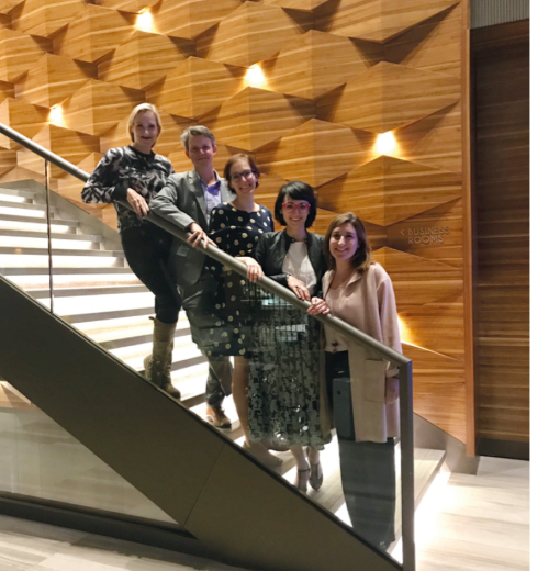
In tune with young radiologists needs, and in order to increase the interest for breast imaging among residents and fellows, in 2015 a platform for breast radiologists and breast imaging researchers younger than 40 years was initiated. The EUSOBI Young Club (EYC) was created as a non-political and non-profit network of younger professionals with the common interest to support and spread the knowledge of breast imaging. It provides a highly accessible platform for enthusiastic young researchers, residents and radiologists and facilitates interaction with acknowledged experts in the field.
The EYC embraces the opportunities offered by social media, since these media platforms have drastically changed how people communicate and how organisations reach their consumers. Young professionals are therefore more easily engaged using these communication channels. Through the EYC, EUSOBI is now active on Facebook, Twitter and Slack to connect breast imagers, breast cancer patients and other physicians in the field of breast care. On our online channels, followers may expect updates on ongoing research highlighting exciting new breast cancer imaging tools, and updates on existing breast imaging standards. In 2018, at the EUSOBI Annual Scientific meeting which will be held in Athens (Greece), the EYC also organises an event specifically for young breast imagers focusing on internal job opportunities and specific challenges that young radiologists currently face.


