HealthManagement, Volume 17 - Issue 3, 2017
 PRINT OPTIMISED
Anaesthetists working in perioperative medicine have increasingly taken a whole body approach to patient evaluation known as TUBE – Total Ultrasound Body Examination – thanks to the development of point-of-care ultrasound. Dr Christophe Aveline, Consultant Anaesthetist in critical care and surgery at the Sévigné Private hospital in Rennes, is an advocate of TUBE and works closely on its adoption with the European Society of Regional Anaesthesia (ESRA) and the French Society of Anaesthesia and Resuscitation (Société Française d’Anesthésie et de Réanimation SFAR). He discusses the importance of the TUBE approach for anaesthetists who specialise in patient care in surgical and emergency procedures, as well as intensive care.
The Sévigné Private hospital in Rennes offers oncology, haematology, surgical and emergency services for more than 20,000 patients a year, covering trauma and orthopaedics, thoracic, abdominal, urological, ophthalmic, reconstructive (breast) and ENT surgery. It has a mixed surgical and medical perioperative department, with eight beds dedicated to postoperative continuous care, plus eight in intensive care. These are almost exclusively used for patients who have undergone complex thoracic, abdominal and orthopaedic or spinal procedures, and trauma patients.
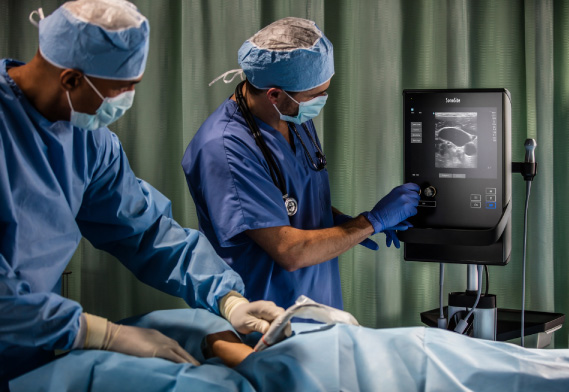
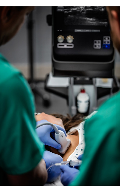
The site’s team of 15 anaesthetists take daily charge of patients in the emergency department, intensive care and surgical theatres, as well as those needing perioperative consultations. We routinely use ultrasound for venous access, regional anaesthesia, and cardiac and pulmonary evaluations, performing upwards of 40 procedures every day on our point-of-care equipment; it is an essential tool for today’s anaesthetist, helping them to care for patients and follow recent recommendations.
Historically, anaesthetists first began to use point-of-care ultrasound for guiding regional anaesthesia and, in the last five years, they have become knowledgeable and skilful in this different approach to performing and interpreting scans at the patients’ bedside. We now use point-of-care ultrasound to answer complex questions about our patients. For example, when a patient is admitted to the emergency department with a fractured hip, the first use of ultrasound is to treat the pain with a guided femoral nerve block, then a full evaluation looks at the lungs, the heart, volaemia, and for a distended bladder. This kind of injury is often seen in older patients, who may have underlying comorbidities, such as cardiac or pulmonary problems, or are on many different treatments. Looking for major cardiorespiratory dysfunction reduces the risk of medical complications related to or aggravated by the injury, and optimises patient care under anaesthesia. Similarly, in trauma patients, pointof- care ultrasound allows us to look for intra-abdominal, retro-peritoneal and pleural effusions, or pneumothorax, to evaluate the presence of pulmonary oedema and pneumopathy. Evaluating blood volume guides vascular filling in cases of hypovolaemia. This whole body approach, known as TUBE , delivers a strategy for evaluating lesions and analgesia. Ultrasound also gives us information on the presence of a distended bladder that may need to be drained.
Guiding regional anaesthesia is a major application of point-of-care ultrasound. Other applications have also been developed, such as guiding difficult intubations, or selective intubations for thoracic surgery. In general, we only use regional anaesthesia in isolation for localised limb procedures, although it may sometimes be used to treat other surgical procedures (breast, thoracic, abdominal: thoracic and abdominal wall, epidural space, paravertebral blocks) or chronic pain in hospitalised patients following thoracotomy or other major surgery. The combined use of regional and general anaesthesia is common in thoracic and abdominal surgery, for example, for investigating the perimedullary space. This last point is important to help avoid misplacement of punctures using a ‘blind’ approach; ultrasound allows a reduction in the number of punctures. 65% of our patients have ambulatory surgery, the others are urgent cases or need to stay in hospital because of a major procedure, or due to the presence of several comorbidities. Long-term perineal catheter placement may be an effective strategy for testing or reducing chronic pain. Ultrasound makes it possible to optimise the positioning of the catheter and to secure it with respect to the adjacent vascular structures.
Young doctors in training during their six month internship with our team participate in ultrasound demonstrations to improve the basic techniques learned during their university studies. Our role as experienced anaesthetists is to supervise and help them to apply this knowledge in real-life situations. Our teaching is not limited to regional anaesthesia, and includes all point-of-care procedures. We also teach them the order in which to follow a procedure so that nothing is missed or goes wrong.
The three FUJIFILM SonoSite S-Nerve™ devices are used daily and valued for their simplicity of use and quick start-up, their mobility, their robustness and their efficiency, as well as for the 2D modality, time-movements and Doppler function. We are often called to go to emergency or intensive care to evaluate a patient who is difficult to mobilise, and the devices give a real ‘point-of-care’ solution. They are ideally suited to the TUBE protocols we perform; we are convinced that this approach will rapidly become a medical necessity. Currently more than 50% of our patients benefit from point-of-care ultrasound as part of a general clinical evaluation.