HealthManagement, Volume 16 - Issue 4, 2016
 PRINT OPTIMISED
West Suffolk Hospital is a district general hospital located in a 19-hectare parkland site on the outskirts of Bury St Edmunds. The hospital has a strong focus on ultrasound training, and its intensive care trainees have the opportunity to develop their skills in point-of- care cardiac and lung ultrasound using state-of-the-art equipment, as consultant anaesthetist Dr Kaushik Bhowmick explained.
My interest in ultrasound began many years ago in India, when a prominent intensivist from a renowned hospital visited where I was working to demonstrate the importance of cardiac output monitoring using echocardiography. I later came to the UK to further my medical career and went on a few ultrasound courses to improve my knowledge of this modality. However, it’s not until you use ultrasound in your everyday practice that you really develop your practical skills and, when I came to West Suffolk to do my intensive care training, I took advantage of the department’s pocket ultrasound system, using it as much as possible.
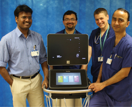
At West Suffolk, we have a 10-bed IT U with 11 consultants. Around two thirds of our cases are medical – COPD , asthma, oncology, haematology, GI – and the remainder are general surgical patients. Like any other intensive care unit, we perform in-house echocardiograms, and are very positive about the use of point-of-care ultrasound. However, when I joined the hospital as a consultant anaesthetist in 2013, the IT U did not have an intensive care-specific ultrasound system that could be used to perform critical care ultrasonography, including echocardiography. With the department’s support, we made a business case and, with some definite goals in mind, trialled several instruments before choosing a FUJIFILM SonoSite X-Porte®.
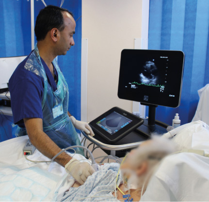
My own main focus is in point-of-care cardiac scanning, while my colleague Dr Vijayakumar Gopal is particularly interested in lung ultrasound. We needed a system that would be suitable for both of these applications, so that we could provide the best service possible for our patients. The first consideration was focused echocardiography, which allows us to quickly assess a patient, establish their fluid status and treat accordingly. Diagnostically though, there are many situations where ultrasound can be helpful, for example, answering the question of whether a patient with hypotension of unknown cause is suffering from a pleural effusion, which can have the same effect as a pericardial effusion in certain circumstances. Ultrasound also helps us to monitor cardiac output as we can see how the heart is contracting and, in the case of a cardiac arrest, we can see whether or not there is any flow within the heart chamber, or possibly detect any heart defect. In an intensive care environment, point-of-care ultrasound is particularly beneficial when an echocardiogram is urgently needed, perhaps in the middle of the night when services are limited. Similarly, an echocardiogram is always requested before a respiratory patient is accepted for extracorporeal membrane oxygenation (ECMO), so that any contraindications for a particular type of ECMO (VV or VA) have been diagnosed.
 |  |
Lung ultrasound is just as valuable as that for cardiac applications and, in many cases, can avoid the need for a chest X-ray. It allows you to see whether or not there is a pleural effusion that should be drained, and also to instantaneously detect pulmonary consolidation seen in patients with pneumonia. This can, on occasions, be quite critical because, with ultrasound, objective data is available immediately, allowing the correct treatment to be started straight away, whereas X-ray tends to lag behind and there may be a delay before a consolidation has developed sufficiently to show up.
The X-Porte ultrasound system is ideal for these purposes and also serves as an excellent teaching device for our intensive care trainees. We place a strong emphasis on training and, in fact, West Suffolk is the first district general hospital in the eastern region to offer a FICE training course. Good ultrasound teaching is very attractive to advanced trainees who rotate within the university hospitals – either Addenbrooke’s in Cambridge, or the Norfolk and Norwich – before spending six months in one of the smaller hospitals in the region. We are effectively competing with other hospitals for these trainees, who are a real asset to IT U, and we need to show that we have something different to offer them. The excellent ultrasound equipment and training opportunities we provide have had a big impact on this.
For all trainees, practice is the key to developing their skills and the X-Porte can really help in this respect. For example, there may be occasions when a trainee cannot get the view they want, but their tutor or a consultant may not be around to ask for advice. The X-Porte allows them to look at the multimedia demonstration module already embedded within the machine. Interpretation of the data is of course even more important, and using the X-Porte for seminars is perfect for showing the trainees different images from real patients, and talking them through a full range of case studies and possible scenarios. In addition to this targeted education for trainees, we are very keen for all our consultants in IT U to gain FICE accreditation. It’s something that I am actively promoting, because if the responsibility for cardiac and lung ultrasound lies with just one or two people it can be difficult to get an assessment done quickly, particularly during the night. The days are long gone when it was acceptable for only selected individuals to use a particular technology. Everybody should learn to use ultrasound, as the more people who can use it, the better it will be for our patients.