The integration of artificial intelligence into breast cancer screening holds promise for addressing workforce shortages and enhancing diagnostic accuracy. While previous studies have demonstrated that AI decision support can improve sensitivity without compromising specificity or reading time, its influence on radiologists’ visual search strategies has not been thoroughly explored. Understanding how AI affects visual search behaviour is critical to optimising its clinical utility. A recent study aimed to investigate the impact of an FDA-approved AI decision support system on radiologist performance and eye movement patterns during screening mammography interpretation.
Improved Detection Performance without Added Time
The study evaluated 12 experienced Dutch screening radiologists who reviewed 150 mammographic examinations with and without AI support. The AI system provided a risk category and optional region markers highlighting areas of concern. When aided by AI, radiologists achieved a higher average area under the receiver operating characteristic curve (AUC) compared to unaided reading (0.97 vs 0.93), indicating improved overall diagnostic accuracy. Subgroup analyses confirmed that this improvement persisted across various lesion types, tumour sizes, breast densities and radiologist experience levels.
Must Read: Cost-Efficient Mammography: Sharing Tasks Between AI and Radiologists
Despite this enhancement, there was no statistically significant difference in average reading times between AI-supported and unaided interpretations (30.8 vs 29.4 seconds), suggesting that the AI tool improved performance without imposing additional time burdens. Moreover, sensitivity and specificity were comparable between conditions, with a trend towards higher sensitivity in very high-risk cases and higher specificity in low-risk ones when using AI support.
Targeted Visual Attention and Streamlined Search
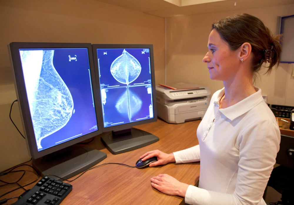
Eye-tracking analysis revealed important behavioural changes when radiologists used AI assistance. The overall area of the breast covered by visual fixations was significantly lower during AI-supported readings (9.5% vs 11.1%), while the time spent fixating within lesion regions increased (5.4 vs 4.4 seconds). These findings suggest that radiologists adopted a more focused search pattern, concentrating more on AI-identified regions and less on non-suspicious areas.
This shift in visual strategy did not affect the time taken to first fixate on a lesion, which remained similar across conditions. Interestingly, radiologists tended to review more of the breast area before activating AI markers and then shifted to a concentrated focus on AI-flagged regions. This phased search approach may balance independent visual assessment with AI-guided confirmation, helping mitigate the risk of automation bias. These outcomes align with prior research showing that computer-aided detection can lead to more restricted but effective search patterns.
Error Types and Workflow Implications
Despite improved lesion-focused fixation, the analysis did not find statistically significant differences in search, recognition or decision errors between the two conditions. This suggests that while AI support helps direct attention more efficiently, it does not necessarily change the underlying error types leading to missed cancers. Importantly, reading time decreased for examinations categorised by AI as low-risk and increased for those assessed as medium-high risk. This redistribution of effort implies that AI can help radiologists allocate time more effectively based on the predicted malignancy likelihood.
In real-world practice, where most screening cases are low risk, such efficiency gains could translate into reduced overall workload without compromising diagnostic accuracy. However, the reduced visual coverage observed with AI raises questions about the possibility of overlooking lesions that AI might miss, highlighting the continued need for radiologists to maintain a critical and comprehensive approach to image interpretation.
The study provides compelling evidence that AI decision support enhances radiologists’ performance in screening mammography by improving diagnostic accuracy and guiding more efficient visual search, without increasing reading time. Radiologists spent more time examining suspicious regions and less on the rest of the breast, adopting a more targeted and potentially effective strategy. However, the unchanged error rates and visual coverage caution against overreliance on AI, emphasising the need for high AI accuracy and informed human oversight. Future research should evaluate these findings in routine clinical practice and explore optimal strategies for presenting AI information to support radiologist decision-making without diminishing their autonomy.
Source: Radiology
Image Credit: iStock


