


Recent advances in noninvasive cardiac imaging technologies have altered the diagnostic approach to patients with coronary artery disease (CAD). Improvements in hardware and software as well as the introduction of new hybrid scanners have allowed the performance of hybrid imaging in ischaemic heart disease, which is now widely adopted and also implemented in international guidelines, according to a review article in the journal Revista Española de Cardiología.
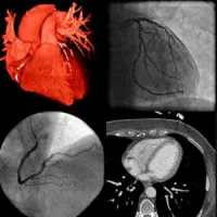
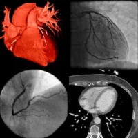
Hybrid imaging for ischaemic heart disease, the article explains, refers to the fusion of information from a single or usually from multiple cardiovascular imaging modalities enabling synergistic assessment of the presence, the extent, and the severity of coronary atherosclerotic disease along with the haemodynamic significance of lesions and/or with evaluation of the myocardial function.
"Physicians have long mentally fused information from different modalities that, combined with the clinical status of the patient, has allowed them to decide on the diagnostic approach and the most appropriate therapeutic management," the article says. "In the era of multimodality imaging, the advent of hybrid imaging has eased this mental fusion of the wealth of information available."
Coronary artery anatomy and coronary plaque morphology, for example, is readily provided by coronary computed tomography angiography (CCTA) while myocardial perfusion imaging (MPI), including single-photon emission computed tomography (SPECT), positron emission tomography (PET) and cardiac magnetic resonance imaging (MRI), can identify regional myocardial perfusion defects, thereby indicating haemodynamically significant lesions.
"The combination of this information could minimise the percentage of false negative and false positive results of each modality, thereby increasing our diagnostic accuracy," authors Andreas A. Giannopoulos, MD and Oliver Gaemperli, MD, PhD, both from the Department of Nuclear Medicine, Cardiac Imaging, University Hospital Zurich (Switzerland) point out.
In principle, most hybrid approaches require that the patient is sequentially imaged in different scanners or in the same scanner. However, newer systems such as PET/cardiac MRI also allow simultaneous acquisitions of signal with both modalities and open new avenues for research and clinical applications.
Furthermore, most of the novel SPECT/CT systems provide newer image reconstruction techniques, such as iterative reconstruction algorithms that allow for improved count sensitivity and enhance image quality.
"Besides high-end hybrid modalities, the combination of native coronary artery calcium scoring-CT scans with SPECT MPI represents a simpler hybrid approach that is easier to implement into clinical practice, since many centres already perform coronary artery calcium scanning on a large scale either as part of a formal CCTA or as a screening routine examination," the authors note. "Even older-generation scanners such as 64-slice CCTAs have been shown to have good agreement with invasive coronary angiography and this agreement was independent of the calcium score."
The article also highlights increased interest in these novel hybrid methods: coronary computed tomography angiography-derived fractional flow reserve (CT-FFR) and computed tomography perfusion. The authors explain: "Computed tomography perfusion and CT-FFR are most likely here to stay, and will enjoy their position in the diagnostic approach of the patient with ischemic cardiac disease, once the current limitations for its widespread adoption, including high radiation for CTP and high costs for CT-FFR, are addressed."
Recently, the fusion of CCTA and 3D speckle-tracking stress and rest echocardiography has gained attention. Like the fusion of other modalities with CCTA, hybrid CCTA/3D echocardiography allows direct visualisation of the coronary arteries and myocardial strain in the subtended territories, according to the article.
"Hybrid cardiac imaging has entered the clinical arena and is increasingly being implemented in the diagnostic approach of patients with ischaemic cardiac disease. Undoubtedly, more studies are warranted to assess all clinical aspects such as the effect on management decisions, improvement of outcomes, and cost/effectiveness that would justify the increased radiation exposure from several currently-available hybrid approaches," Drs. Giannopoulos and Gaemperli conclude.
Source: Revista Española de Cardiología
Image Credit: Pixabay
References:
Giannopoulos AA, Gaemperli O (2018) Hybrid Imaging in Ischemic Heart Disease. Rev Esp Cardiol. May 2018; 71(5):382–390 https://doi.org/10.1016/j.rec.2017.11.023
Latest Articles
cardiac imaging, ischaemic heart disease, hybrid imaging
Recent advances in noninvasive cardiac imaging technologies have altered the diagnostic approach to patients with coronary artery disease (CAD). Improvements in hardware and software as well as the introduction of new hybrid scanners have allowed the perf