A team of cardiologists at UCLA used a minimally invasive approach to replace a heart valve of a patient whose unique heart anatomy made conventional open-heart surgery too risky.
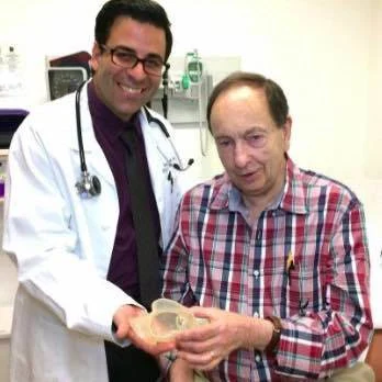
The team printed a 3-D model of Richard Whitaker’s heart to determine whether a replacement valve would align and fit with his unusually large pulmonary arteries.
The 3-D model was created using a CT scan of Whitaker’s heart. It was made with a silicon-like material with properties similar to tissues and other structures of the heart. The team specified what areas of the model would be made from hard material to simulate calcium deposits and which would be made from softer material to represent heart muscle and vascular tissue.
The doctors then guided a mesh-like stent into place so that it would hold the valve and guided the packaged valve into the model. When the valve was in, the doctors opened it to find that it was a perfect fit.
The real procedure was then performed on Whitaker. The doctors guided the valve and stent up to his heart through a small vein in the groin using a catheter. Once the valve was placed and deployed, it started to work. The patient was able to go home only after four days of the procedure.
Dr. Jamil Aboulhosn, director of the Ahmanson/UCLA Adult Congenital Heart Disease Center and the Streisand/American Heart Association Endowed Chair in the Division of Cardiology at the David Geffen School of Medicine at UCLA explains that technologies such as 3-D printing and advances in minimally invasive procedures are making it possible for congenital heart patients to lead full, normal lives.
"The models created from 3-D printing are becoming more and more realistic," he said. "Even now, they allow us to predict which strategies we can use and in which patients. In the future, they will be used to not only simulate procedures but also allow more rapid development of new devices to help individual patients.”
Source: University of California, Los Angeles (UCLA), Health Sciences
Image Credit: University of California, Los Angeles (UCLA), Health Sciences


