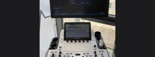
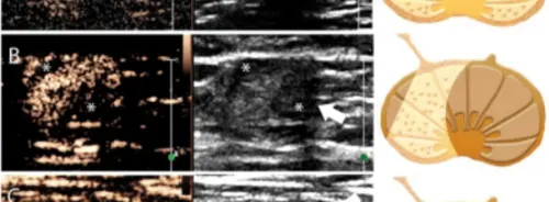
Temporal artery ultrasound is a promising diagnostic tool for giant cell arteritis (GCA), offering a non-invasive alternative to traditional temporal artery biopsy (TAB). A recent prospective study led by Guillaume Denis, MD, from the Centre Hospitalier de Rochefort, France, sheds light on its efficacy and implications. The study, conducted across six French hospitals from August 2016 to February 2020, enrolled 165 patients exhibiting high clinical suspicion of GCA. Criteria included age over 50, elevated C-reactive protein levels, and indicative clinical or general signs of GCA, along with visible large-vessel vasculitis on imaging. All participants underwent temporal artery ultrasound within one week of corticosteroid therapy initiation, with the procedure occurring within a day of specialist consultation.
Diagnostic Outcomes and Challenges in Temporal Artery Ultrasound for GCA
Results demonstrated that temporal artery ultrasound identified GCA in 44% of cases, affirming its diagnostic reliability. Patients with positive ultrasounds exhibited bilateral temporal halo signs, indicating vasculitis. Subsequent confirmation at one month and up to two years of follow-up validated these diagnoses. For patients with negative ultrasounds, 30% were later diagnosed via TAB, underlining the importance of multimodal diagnostic approaches. While ultrasound’s sensitivity and specificity remain high, concerns persist regarding operator dependence and evolving techniques. Furthermore, 25% of GCA cases went undetected by both ultrasound and TAB, highlighting diagnostic challenges. Dr. Mark Matza of Massachusetts General Hospital underscores the need for caution, emphasising the variability in ultrasound performance and the possibility of missed diagnoses.
Advantages and Considerations in Temporal Artery Ultrasound
Despite these limitations, ultrasound’s advantages are evident. Its non-invasive nature reduces patient discomfort and eliminates biopsy-related risks. Moreover, ultrasound’s accessibility and immediacy make it a valuable tool for initial assessment, potentially streamlining diagnostic workflows and reducing costs. Dr. Minna Kohler, also from Massachusetts General Hospital, underscores the importance of multimodality imaging, balancing ultrasound’s strengths with complementary techniques like CTA, MRA, and PET/CT for comprehensive evaluation.
Reevaluating Diagnostic Approaches to GCA
The study’s findings prompt a reevaluation of current diagnostic paradigms. While guidelines favour TAB, ultrasound’s growing acceptance in Europe suggests a paradigm shift towards non-invasive methods. However, regional disparities persist, with the U.S. lagging in ultrasound utilisation due to limited expertise. Dr. Kohler advocates for a holistic approach, leveraging ultrasound’s advantages alongside other modalities to enhance diagnostic accuracy.
Temporal artery ultrasound emerges as a promising diagnostic tool for GCA, offering a non-invasive and efficient alternative to TAB. While challenges remain, its potential to streamline diagnostics and reduce procedural risks warrants further exploration. As diagnostic practices evolve, a multifaceted approach integrating clinical intuition and advanced imaging techniques promises to enhance patient care in diagnosing GCA.
Source: Annals of Internal Medicine
Image Credit: iStock


