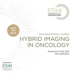
ESHI Educational Course: Hybrid Imaging in Oncology
Start:
Thu, 14 Dec 2017, 09:00
End:
Sun, 15 Oct 2017, 18:00
Website:
Venue:
Exhibit
Symposia
Workshops
Organiser
Sponsor
COURSE DESCRIPTION
This course is aimed at last-year residents, general radiologists, nuclear medicine physicians and oncologists who want to update their knowledge on state-of-the-art hybrid medical imaging of cancer, with a focus on the latest technological advances and clinical applications. No previous practical experience with the subject is required.
The expert faculty includes nuclear medicine physicians and radiologists, as well as physicists and radiopharmacists, who will provide a series of lectures describing imaging pathways for oncological diagnosis, follow-up and treatment response assessment. Lectures will be followed by interactive workshops, presented jointly by faculty specialists..
Learning objectives
- to learn the principles of hybrid medical imaging
- to learn the key clinical questions at different points in the patient’s journey
- to understand the indications, limitations and comparative merits of each part in hybrid medical imaging in a wide range of oncologic conditions, including lymphoma, prostate cancer, gynaecological cancers, as well as brain tumors
- to appreciate the complementary roles of structural and functional/molecular imaging in cancer management
- to understand the importance of standardized protocols for hybrid imaging, and the clinical impact of structured reporting
- to understand how information derived from imaging guides patient selection for treatment and supports individualised treat
You can register to this course by completing the online form on our website. Please note that you are only fully registered after the ESHI office has officially confirmed your registration.
Early fees apply for registrations until November 2, 2017. After this date, the late fees apply.
REGISTRATION FEES
Early fee* | Late fee | |
Non-members | EUR 400 | EUR 450 |
ESHI members | EUR 300 | EUR 350 |
ESHI members in training | EUR 200 | EUR 250 |
*Early fee applies until November 2, 2017 | ||
Thursday, December 14, 2017 | |
09:00-12:00 | Optional site visit at Medical University Vienna: PET/CT, SPECT/CT, PET/MR Join a guided tour through the Radiology and Nuclear Medicine department at the Medical University Vienna and experience the daily work on PET/CT, SPECT/CT and PET/MR Scanners from admission of a patient to diagnosis. There
will be 3 tours à 45 minutes. |
12:00-13:00 | Registration |
13:00-14:00 | Introduction to Hybrid Imaging |
14:00-15:00 | Lymphoma |
15:00-15:30 | Coffee Break |
15:30-17:00 | Hands on Sessions |
Friday, December 15, 2017 | |
08:30-09:30 | Hybrid Imaging in
Neuro-Oncology |
09:30-10:30 | Hybrid Imaging in Prostate
Cancer |
10:30-11:00 | Coffee Break |
11:00-12:30 | Hands on Sessions |
12:30-13:30 | Lunch Break |
13:30-14:30 | Hybrid Imaging in
Gynaecologic Cancer |
14:30-15:30 | Standardized Protocols and
Structured Reporting |
15:30-16:00 | Coffee Break |
16:00-17:30 | Hands on Sessions |
17:30-18:00 | Final Test (multiple choice questions) |
18:00 | Farewell and Certificates |
More events
Thu, 6 Jun 2024 - Sat, 8 Jun 2024

Mon, 10 Jun 2024 - Wed, 12 Jun 2024
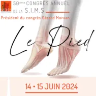
Fri, 14 Jun 2024 - Sat, 15 Jun 2024
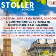
Sat, 15 Jun 2024 - Sun, 16 Jun 2024
Thu, 20 Jun 2024 - Sat, 22 Jun 2024
Sat, 22 Jun 2024 - Wed, 26 Jun 2024


