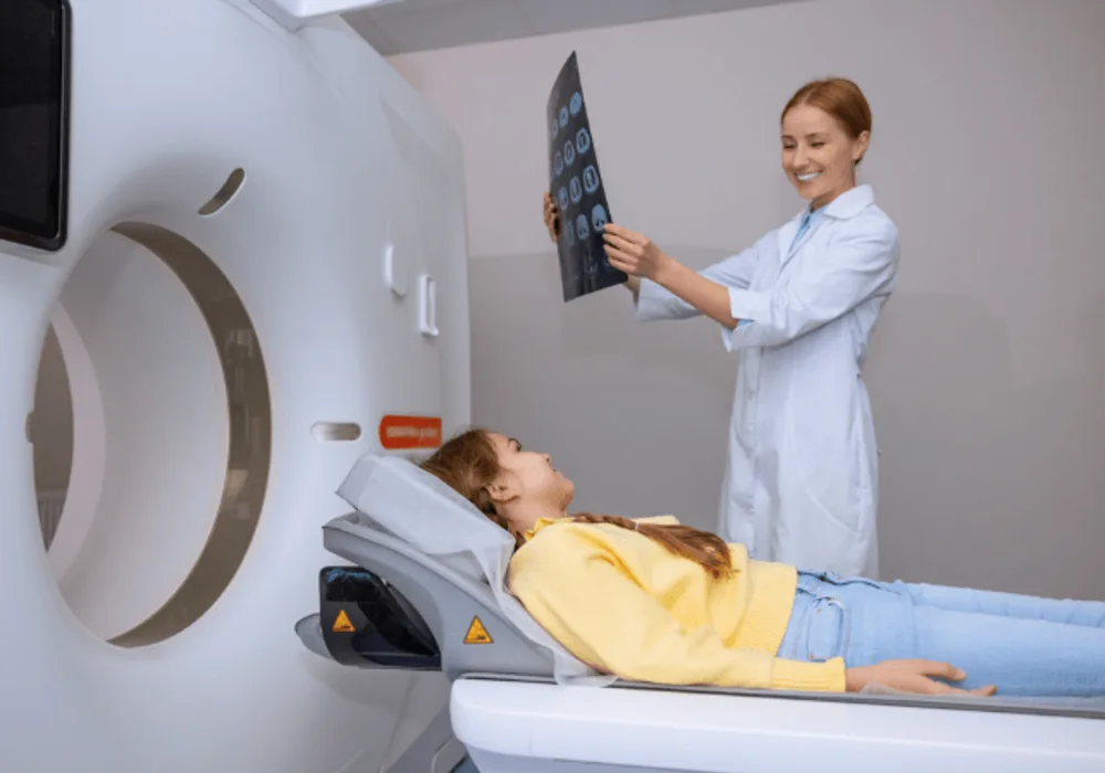
Photon-counting detector CT (PCD-CT) is a cutting-edge advancement in CT imaging technology that addresses limitations of conventional methods. It enhances visualisation of small anatomical structures and may enable higher precision with lower radiation exposure. The unique features of PCD-CT require updated scanning protocols for various diagnostic tasks. Early studies in paediatric populations show promising results in lung, abdomen, and bone imaging, but research on cardiac and vascular imaging is limited. A recent review published in Radiology Advances provides an overview of PCD-CT principles, technical considerations, and its potential benefits in paediatric lung, cardiac, and vascular imaging.
Breaking Boundaries in CT Imaging
Current energy-integrating detector CT (EID-CT) relies on solid-state scintillator crystals to convert x-ray photons into visible light, which is then transformed into an electrical signal. However, this process has limitations, including reduced spatial resolution due to septa between detector elements, underweighting of lower energy photons affecting image contrast, increased electronic noise at lower doses, and limitations in dual-energy CT (DECT) imaging. In contrast, photon-counting detector CT (PCD-CT) directly converts x-ray photons into electrical signals using semiconductor materials. PCD-CT offers advantages such as smaller detector elements, higher spatial resolution, improved iodine contrast-to-noise ratio, reduced electronic noise, and enhanced spectral imaging capabilities. These technical advancements make PCD-CT a promising option for various diagnostic tasks.
Enhancing Safety and Efficiency
Safety concerns in paediatric CT imaging focus on minimising radiation dose and contrast media usage. Photon-counting detector CT (PCD-CT) enhances dose efficiency by eliminating septa between detector elements and reducing electronic noise. It employs tin filtration to remove low-energy photons, decreasing radiation exposure without compromising image quality. PCD-CT also optimises contrast media usage by weighting low-energy photons more heavily, increasing iodine signal-to-noise ratio. Additionally, it generates virtual monoenergetic images at lower photon energies, allowing for reduced contrast volumes while maintaining image quality. Studies suggest PCD-CT can achieve non-inferior image quality with a 25% reduction in contrast media volume compared to conventional CT. These advancements are particularly beneficial for paediatric patients, including those with renal impairment or limited venous access.
PCD-CT Advances in Thoracic Imaging
Photon-counting detector CT (PCD-CT) offers significant clinical benefits in thoracic imaging, particularly in evaluating parenchymal lung disease, lung nodules, and mediastinal masses. For parenchymal lung disease, PCD-CT provides improved visualization of fine structures in the lung parenchyma compared to conventional CT (EID-CT), due to thinner slices and larger matrix sizes. Studies demonstrate enhanced detection of bronchial walls and pathologies such as honeycombing, fibrosis, and emphysema with PCD-CT. Additionally, PCD-CT enables greater dose reductions while maintaining diagnostic image quality compared to EID-CT. In lung nodule characterization, PCD-CT's thinner slices and larger matrix sizes enhance edge definition and shape characterization, potentially impacting management decisions such as metastasectomy. In evaluating mediastinal masses, PCD-CT achieves significant dose reductions while maintaining diagnostic image quality compared to EID-CT. Studies show a reduction in radiation dose by up to 65% with PCD-CT, making it a favourable option for pediatric patients. Overall, PCD-CT improves diagnostic accuracy, enhances image quality, and reduces radiation dose in thoracic imaging, making it a valuable tool in clinical practice.
Cardiovascular Benefits of PCD-CT
In cardiovascular imaging, photon-counting detector CT (PCD-CT) offers notable advantages in evaluating congenital heart lesions, extracardiac vessel anomalies, and stents. For congenital heart disease assessment, PCD-CT provides higher contrast-to-noise ratios compared to conventional CT (EID-CT) with equivalent radiation doses. It enhances visualisation of normal anatomic structures and intracardiac pathology, potentially eliminating the need for further dedicated coronary artery imaging. PCD-CT's higher resolution enables better definition of coronary artery origins and proximal branches. In stent evaluation, PCD-CT overcomes artifacts associated with conventional CT, allowing for more accurate measurement of stent diameters and luminal patency. This capability is particularly valuable for children with obstructive lesions or ductus-dependent circulation. In thoracic vessel assessment, PCD-CT's higher spatial resolution and iodine signal improve characterization of vascular pathologies such as aortic coarctation, patent ductus arteriosus, and pulmonary embolism. It offers better edge and size definition compared to EID-CT, with the potential for dose reduction. Overall, PCD-CT enhances diagnostic accuracy and reduces radiation dose in cardiovascular imaging, making it a valuable tool for paediatric patients with congenital heart disease and vascular anomalies.
Harnessing Spectral CT with PCCT
Spectral CT, facilitated by photon-counting detector CT (PCD-CT), utilizes multiple photon spectra to generate images. PCD-CT uniquely provides spectral information in every scan using a single tube potential, detecting the energy of individual photons. Virtual monoenergetic images (VMIs) at different energy levels are constructed for various diagnostic tasks, such as enhancing iodinated contrast material or decreasing noise levels. Typical energy levels range from 55 keV for angiography to 70 keV for non-contrast CT. Spectral imaging also enables material-specific images like iodine maps and virtual non-contrast images. Iodine maps aid in lesion characterization and treatment response assessment, while virtual non-contrast images help differentiate high-attenuating lesions. Pending FDA approval includes applications like lung perfused blood volume (PBV) and lung vessel analysis, which can contribute to evaluating perfusion in vascular and parenchymal diseases.
Photon-counting detector CT (PCD-CT) shows promise in overcoming limitations of conventional energy-integrating detector CT (EID-CT) in pediatric clinical practice. Its advantages, including superior spatial resolution, iodine contrast, noise properties, and intrinsic spectral imaging, enhance visualization of small physiological and pathological structures, increasing diagnostic confidence in lung and cardiovascular imaging tasks in children. PCD-CT also contributes to radiation dose reduction, crucial in pediatric imaging. Optimizing scanning factors to leverage PCD technology is essential for realizing these benefits. While early experiences are promising, larger studies are needed to assess PCD-CT's capabilities in broader patient cohorts and its impact on diagnostic sensitivity and disease classification in lung and cardiovascular conditions. Future research should explore the effects of deep learning in image reconstruction and noise reduction algorithms on PCD-CT image quality, radiation dose, and diagnostic accuracy. Overall, this technology holds significant potential for advancing pediatric imaging practices.
Source: Radiology Advances
Image Credit: iStock


