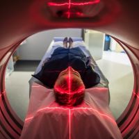
A group of researchers from Italy, led by Dr Paul Summers, from the European Institute of Oncology (IEO), presented results on “Whole-body magnetic resonance imaging: technique, guidelines and key applications.” As more clinicians and patients are requesting a radiation-free technique to deal with cancer screening and management, whole-body magnetic resonance imaging (WB MRI) offers such a solution. This study reviewed its technical basis, current international guidelines for use and key applications.
WB MRI is an imaging method that does not use ionising radiation, giving whole-body (WB) coverage with a core protocol of essential imaging contrasts in less than 40 minutes. Moreover, it can be complemented with sequences to evaluate specific body regions, as needed.
To understand the context, one has to go back to the 1990s, when multiple receiver coils and table movement for multi-station scanning were introduced. This completely changed MRI from segmental to a whole-body imaging modality. Improvements in scanner homogeneity and gradient systems led to the introduction of diffusion-weighted imaging (DWI) whole-body examinations.
During the early 2000s, more and more evidence showed that DWI gave a good diagnostic performance in detecting, characterising and monitoring of therapy for many types of cancer. As the technology improved, for instance accelerated imaging and sequence design, image quality became much better, resulting in WB MRI becoming clinically acceptable as the scan-time was quite rapid.
Often WB MRI is superior to bone scintigraphy and computed tomography in detecting and characterising lesions, evaluating their response to therapy and screening high-risk patients. Also, due to its spatial resolution, high sensitivity, non-invasive nature and absence of ionising radiation, this imaging technique has become the preferred choice of many clinicians.
Moreover, international guidelines recommend that WB MRI is used to manage patients with multiple myeloma, prostate cancer, melanoma and individuals with certain cancer predisposition syndromes. It is also increasingly used for metastatic breast cancer, ovarian cancer and lymphoma.
Over the last 20 years, technological advances made in scanner hardware and imaging techniques have led to WB MRI that provides a core clinical protocol for WB imaging in about 40 minutes. The future bodes well as artificial intelligence and imaging strategy continue to develop and expand to other applications in oncology. However, it is essential that reporting and acquisition are standardised to reduce the variations in diagnostic performance.
Source: NCBI
Image credit: iStock


